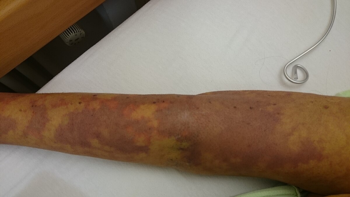
Sepsis, often called the “silent killer,” is a global health crisis that claims millions of lives each year. Despite its alarming impact, early detection remains one of the most significant challenges in managing this condition. Recent advancements by Canadian researchers have brought fresh hope, as they unveiled a groundbreaking, non-invasive imaging technique that could revolutionize how sepsis is detected in its early stages.
Sepsis occurs when the body’s response to an infection spirals out of control, causing widespread inflammation and potentially leading to life-threatening organ failure. It’s not a disease in itself but a severe complication that can result from infections such as pneumonia, urinary tract infections, or even minor wounds.
The condition progresses rapidly, making early diagnosis critical. Yet, due to its complex nature, clinicians often face difficulty recognizing sepsis in its nascent stages. This delay can lead to devastating consequences, including irreversible organ damage and death.
Researchers from Western University in Ontario, Canada, have introduced a promising imaging strategy that could change the way sepsis is diagnosed. Their findings, published in The FASEB Journal, highlight the potential of two non-invasive imaging techniques to detect early signs of sepsis by analysing blood flow in skeletal muscles.
Unlike traditional diagnostic methods, which rely heavily on identifying changes in vital organs like the brain or heart, this new approach focuses on skeletal muscle microcirculation. According to the researchers, skeletal muscle may show signs of compromised blood flow earlier than vital organs, making it an ideal target for early detection.
The team employed two advanced imaging methods:
1. Hyperspectral Near-Infrared Spectroscopy (HNIRS): This technology measures oxygen levels in tissues by analysing how light interacts with them. It provides a detailed view of blood flow and tissue oxygenation, helping identify abnormalities before they escalate.
2. Diffuse Correlation Spectroscopy (DCS): DCS assesses microvascular blood flow by analysing the movement of red blood cells. It’s a non-invasive, bedside-friendly method that offers real-time insights into microcirculatory function.
These techniques, already used to monitor tissue conditions in critical care, showed remarkable potential during experiments conducted on rodents. The researchers observed changes in skeletal muscle blood flow even before damage was detectable in vital organs like the brain.
Why Skeletal Muscle?
The study revealed that skeletal muscle could serve as an early warning system for sepsis. While the brain and other vital organs are somewhat protected in the early stages, skeletal muscle microcirculation is more vulnerable. By targeting this early indicator, clinicians could intervene before the condition escalates, potentially saving countless lives.
Currently, sepsis management relies on early administration of antibiotics and vasopressors to combat infection and maintain blood pressure. However, these treatments are often initiated only after clinical signs of sepsis become apparent, which may be too late for some patients.
The global healthcare community faces a pressing need for accessible, affordable, and non-invasive diagnostic tools. The imaging techniques developed by the Canadian researchers could fill this gap, offering a reliable way to detect sepsis in its earliest stages.
Sepsis disproportionately affects vulnerable populations in low-resource settings, where healthcare infrastructure is limited. The accessibility and affordability of these imaging technologies could make a significant difference in such regions. Portable, non-invasive devices could enable healthcare providers to screen patients for sepsis in rural clinics or disaster zones, where early diagnosis is often a matter of life and death.
The researchers plan to test their imaging techniques on patients in intensive care units (ICUs). They hope to refine these methods for clinical use by monitoring microcirculatory function in real-time. If successful, this technology could pave the way for a new standard in sepsis care, enabling earlier intervention and improved outcomes.
“Sepsis is a leading cause of death worldwide,” said Rasa Eskandari, a doctoral candidate at Western University and co-corresponding author of the study. “Early recognition can significantly improve outcomes and save lives. Our team is committed to developing accessible technologies for timely sepsis detection.”
Sepsis is a complex and multifaceted condition that requires a coordinated effort to combat. Beyond technological advancements, there is a need for:
- Public Awareness: Many people are unaware of the symptoms and severity of sepsis. Educational campaigns can help individuals recognize early signs and seek medical attention promptly.
- Policy Changes: Governments and healthcare organizations must prioritize funding for research and development of diagnostic tools. Policies should also focus on improving access to healthcare in underserved regions.
- Clinical Training: Healthcare professionals need regular training on the latest diagnostic and treatment protocols for sepsis. Early recognition by clinicians can make a critical difference in patient outcomes.
The development of non-invasive imaging techniques marks a significant step forward in the fight against sepsis. By focusing on early detection, researchers address one of the most challenging aspects of managing this life-threatening condition.
While the journey from lab to clinic is still ongoing, the potential impact of these technologies cannot be overstated. They offer hope not only for improved patient outcomes but also for a future where sepsis is no longer a silent killer.
As healthcare continues to advance, innovations like this remind us of the power of science to save lives and bring hope to those who need it most. By investing in research, raising awareness, and fostering global collaboration, we can move closer to a world where sepsis is detected and treated before it claims another life.

 As healthcare continues to advance, innovations like this remind us of the power of science to save lives and bring hope to those who need it most.


.png)
.png)
.png)
.png)


.jpeg)

.jpeg)


.jpg)


.jpg)

