 Saturday, October 10, 2026
 White Paper: Lung Ultrasound in patients with Coronavirus




Since December 2019, the outbreak of pneumonia caused by a new coronavirus, which was later identified as coronavirus disease 2019 (COVID19), has infected innumerable patients globally according to the situation report of World Health Organization. Lung ultrasound is an important tool for the diagnosis and follow-up of pneumonia in neonates, children, and adults. Recent CT reports demonstrated that most of the lesions were distributed peripherally in the lung, which facilitates detection by lung ultrasound. The lung ultrasound findings of COVID-19 pneumonia and the study of the relationship between the ultrasound findings and clinical severity and the time-course of disease progress were measured to arrive at the conclusion. Bedside lung ultrasound was performed to detect B-lines, lung consolidation, and pleural line abnormalities at 5 areas in each lung. Vascular ultrasound was also performed to detect potential deep vein thrombosis.
A total of 20 patients of COVID-19 pneumonia (12 males and 8 females) were categorized as 4 moderate, 5 severe, and 11 critical cases according to the current diagnosis and treatment program. All patients showed abnormal lung ultrasound findings, including 100% (20) pleural line abnormalities, 100% (20) B-lines, and 50% (10) consolidation. Most of the moderate and severe cases could show both separated B-lines and confluent B-lines during admission. All critical patients showed confluent B-lines, and 18% (2) of them had compact B-lines. Bilateral involvement was observed in all patients. The predominantly involved areas in moderate patients were on the back, i.e., the interscapular and infrascapular areas. For severe and critical patients, all 5 areas could be involved. Consolidations were not detected in moderate cases and distributed mainly on the posterior areas in severe and critical cases. Pleural effusion (18%, 2 cases), pericardial effusion (9%, 1 case), and deep vein thrombosis (64%, 5 cases) were only found in critical patients. (Table 1).
A total of 36 ultrasound examinations were categorized into four groups based on the time interval between onset of symptoms and ultrasound examinations (1st to 4th week). All of the examinations showed abnormal lung ultrasound findings, including 100% (36) pleural line abnormalities, 100% (36) B-lines, and 64% (23) consolidation. The separate B-lines were found more than half of the examinations after the 2nd week. The majority of examinations during the 2nd and 3rd weeks showed confluent B-lines. The involvement of anterior areas with B-lines decreased along with the infected time course. The lateral and back areas were always involved in all stages for B-lines. Consolidations were found in more than half examinations after the 1st week. Most of the consolidation lesions confined within unilateral lung except in the 2nd week. The anterior and lateral areas were not involved in consolidations during the 1st and 4th weeks. Consolidations were always found in the interscapular and infrascapular areas. Pleural effusion was observed across all stages, but only in a few examinations. The deep vein thrombosis can be found since the 1st week and most prevalent in the 4th week (62.5%, 5 cases). (Table 2).
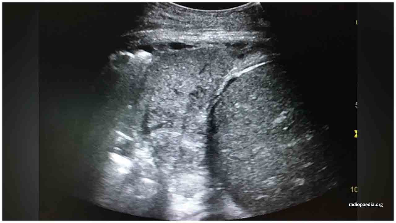
In summary, abnormal lung ultrasound findings, mainly B-lines, consolidation and pleural line abnormalities, were found in patients infected with COVID-19 pneumonia. Bilateral involvement was always observed with predominant distribution in the posterior part of the lungs.
The composition of different density of B-lines and areas of consolidation showed parallel changes with the clinical severity. The extent of disease demonstrated by ultrasound findings seemed to reach the peak in the 2nd week and recovering gradually thereafter. Collectively, lung ultrasound could serve as a valuable tool for the detection and follow-up of lung lesions in COVID-19 pneumonia and also provide supplemental imaging information for current recommended radiological examination, with the advantages of radiation-free, flexibility, and cost-effective.






.jpeg)










.jpg)
.jpg)
.jpg)
.jpg)
.jpg)
.jpg)


.jpg)