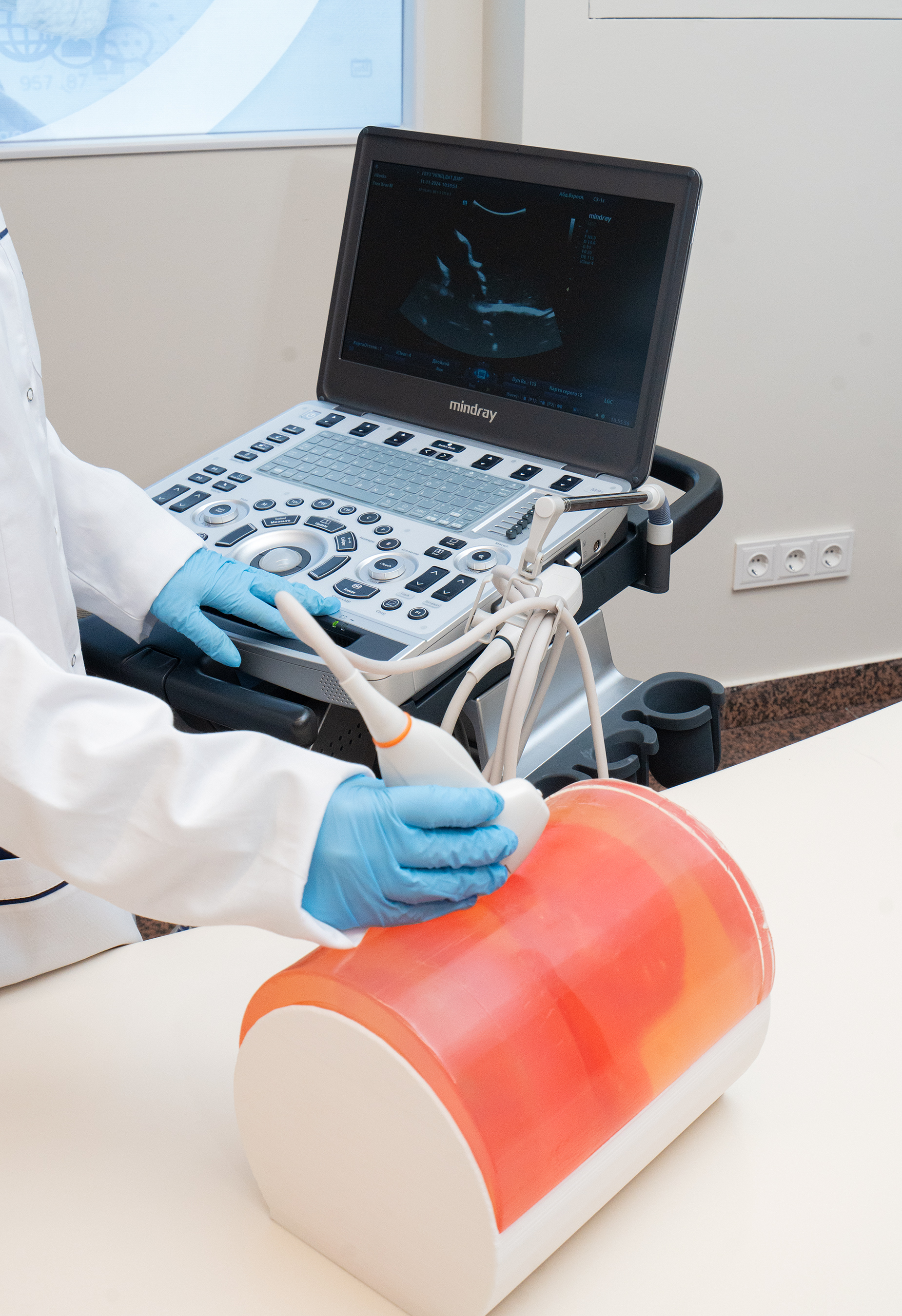
In Moscow, researchers specializing in radiology have successfully created a total of 10 ultrasound-training phantoms, with the latest innovation being a fetal phantom designed for use in ultrasound examinations for expectant mothers. This new fetal phantom accurately replicates the anatomical features of a 20-week-old fetus and produces an ultrasound image that mimics those obtained by specialists during actual scans. This advancement enables students and medical residents to refine their ultrasound examination skills effectively.
Yuri Vasilev, the Chief Consultant for Radiology and CEO of the Center for Diagnostics and Telemedicine of the Moscow Healthcare Department, stated, “Phantoms have become an integral part of medical training today. By practicing on these simulators, physicians can interact confidently with patients, having already honed their skills on these models. This particular development facilitates the training in diagnostic ultrasound. It is the most widely used method for assessing fetal health, capable of identifying up to 80% of congenital disorders at an early stage. The quality of this examination is vital, as it impacts both the mother and the unborn child. Therefore, it is essential that physicians conduct these procedures competently, and the phantom serves to enhance these skills to a level of proficiency.”
The fetal phantom is the tenth model developed by the scientists at the Center for Diagnostics and Telemedicine under the Moscow Healthcare Department. The center has also created models representing various other anatomical features, including the mammary glands, thyroid gland, blood vessels and nerves, liver, facial structures, and the lumbar spine. Additionally, they have designed a phantom for studying blood vessels through the skull, a fetal phantom for MRI, and a model that simulates the human lumbar spine with varying levels of bone mineral density for CT and MRI scans.
“The fetal phantom is a foundational model featuring a representation of amniotic fluid and a healthy fetus at 20 weeks of gestation, which aligns with the timing of the second trimester screening. At this stage, healthcare professionals can assess for potential anomalies and evaluate whether fetal growth aligns with gestational age. This phantom allows for practical training in determining fetal presentation and utilizing navigation sensors to measure key anatomical parameters, including the size and shape of the body, head, and limbs. Its durability ensures repeated use, maintaining its integrity and the quality of ultrasound images without deterioration—a distinct advantage of our domestic models, achieved through the application of innovative materials,” noted Anton Vladzimirsky, Deputy Director for Research at the Center for Diagnostics and Telemedicine.
Since commencing small-scale production of these test objects in 2023, the Center has produced approximately 100 phantoms of various types. Presently, these models are utilized across five medical educational institutions in Russia, including the Russian Medical Academy of Continuing Professional Education, “Association of Interventional Pain Treatment,” the Academy of Medical Education named after F.I. Inozemtsev, Smolensk State Medical University, and the Training Center of the Center for Diagnostics and Telemedicine. The phantoms are also employed to demonstrate equipment from companies such as SonoScape, Samsung, Philips, and Zaslon.
The breast phantom accurately replicates the anatomy of the mammary gland, constructed from silicone-like materials that effectively mimic the ultrasound properties of human tissues. It incorporates modeled pathologies, including cystic formations and tumor foci, enabling future physicians to practice essential ultrasound-guided procedures such as pathology detection and biopsy sampling. Additionally, the phantom features a removable skin for enhanced realism
The phantom that simulates human vessels and nerves includes three modeled vessels and nerves, allowing specialists to hone skills in accessing vessels under ultrasound guidance and performing peripheral nerve blocks for anesthesia. The model facilitates the injection of a simulated anesthetic to verify needle tip placement and practice entire regional anesthesia procedures. The design permits repeated use, as fluids within the phantom are automatically removed.
The thyroid phantom is anatomically shaped like a human neck and encompasses not only the thyroid gland but also includes the trachea, arteries, veins, and cervical spine bones. This model is specifically intended to enhance diagnostic skills related to mass identification and biopsy techniques under ultrasound guidance.
Notably, Russian-made phantoms provide distinct advantages over their imported counterparts, including superior functionality and durability, allowing for repeated use and resulting in lower production costs due to the utilization of Russian materials.
Phantoms have been showcased at international exhibitions and conferences across 11 countries, including Thailand, Brazil, Japan, Turkey, Spain, France, Belarus, Pakistan, China, the United Arab Emirates, and Kazakhstan.
...

 Phantoms have been showcased at international exhibitions and conferences across 11 countries, including Thailand, Brazil, Japan, Turkey, Spain, France, Belarus, Pakistan, China, the United Arab Emirates, and Kazakhstan.


.jpeg)


.jpeg)
.jpeg)
.jpeg)
_(1).jpeg)

_(1)_(1)_(1).jpeg)
.jpeg)
.jpeg)
.jpeg)


.jpeg)
.jpeg)