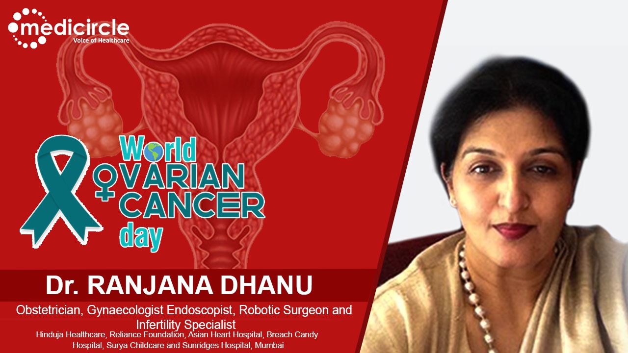
India has the world’s second-highest ovarian cancer incidence. Since the 1980s, there has been a growing drift of ovarian cancer in our country. Medicircle is conducting an exclusive series featuring eminent gynecologists, obstetricians, and oncologists so that people can get direct and more reliable information from them.
Dr. Ranjana Dhanu is an Obstetrician, Gynaecologist, Endoscopist, Robotic Surgeon, and Infertility Specialist. She is practicing at prominent hospitals in Mumbai, including Hinduja Healthcare, Reliance Foundation, Asian Heart Hospital, Breach Candy Hospital, Surya Child Care, and Sunridges Hospital. She is one of the most renowned Minimal Access Surgeons in the country with close to 25 years of experience in which she has added a lot of feathers to her cap and earned immense accolades. She has been invited on numerous occasions to the UK and several countries in South East Asia as an international faculty. Apart from endoscopy, she has catered to several patients for high-risk obstetrics. In addition to her ongoing contribution to society, Dr. Dhanu continues to associate herself with charitable foundations, to address healthcare issues faced by women the world over, and in doing so, ensures that her inherent talent, painstakingly honed over the years, helps her achieve her dream of making quality women’s healthcare a reality.
Who is at risk of having ovarian cancer?
Dr. Dhanu mentions, “Ovarian cancer is not hereditary, rather it has a genetic predisposition. So, if your family members or relatives, especially from the maternal side like mother, grandmother, aunt, sister, or niece have had it, then you fall in the first risk category and there would be a slightly higher risk of ovarian cancer. Also in patients who have had breast cancer, there is a risk. These patients are at risk of having Krukenberg tumors, which are solid ovarian tumors. So short of the long this is how you can encounter ovarian cancer, and you need to be vigilant.”
Ovarian cancer – be thoroughly informed about it
Dr. Dhanu explains, “The uterus is almost like a small chikoo or a pear. On the sides, there are two little ovaries which are like cashew nuts and there are cervix and tubes and the vagina. So understand that these ovaries that are so small on either side, if they start to grow in size from the size of cashew to tomato, or the size of an orange, or the size of an apple or the size of muskmelon, we don't feel that much of discomfort in the abdomen because it's like two small fruits in a shopping bag. The contents adjust themselves around the ovaries.
Now, if you go by the incidence of ovarian cancer, and all the genital cancers in totality, the highest-ranking is that of the breast. But amongst genital cancers, it is the cervix, which is the mouth of the uterus, which is almost 86% of all genital cancers. 8% of cancers are ovarian cancer, but more dreaded. Why more dreaded because there is hardly any presenting symptom in the early part of the disease. When these ovaries start to grow and when they start to become cystic, they do not present themselves with any specific symptoms. A patient with the breast lump will say, “oh I feel a lump or abnormal discharge. A patient with a cervical lesion will say I have abnormal discharge. A patient with uterine cancer will say, I have abnormal intermenstrual bleeding or postmenopausal bleeding”, but if somebody gets ovarian cancer, she may say nothing until the most dreaded thing which is stage 3 wherein it has spread into the abdomen and cause ascites. Ascites are fluid-filled around the ovaries and the bowel. And that is when the woman starts to feel tense in her abdomen and goes for a checkup. Secondly, it can be accidentally diagnosed on routine health checkups.”
Dr. Dhanu further mentions, “There are many times when we have patients with simple ovarian cysts, but they have a malignant component as well. So, understand that anything big starts as something small, you cannot stay with a smaller cyst. So, if you see a cyst, you get vaginal sonography done, based on which it is decided whether it's a simple cyst that is smaller than 4 centimeters or bigger than that. We follow it up immediately after the menstruating patient’s next menses to see whether that's regressing, for any reason. If it's increasing in size, certain tests are done to see whether there is any malignant potential in those cysts.
The nature of the cyst is extremely important. So, we have tests like CA-125, Roma index, CEA, AFP, HE4. These are all advanced tests, which tell us exactly what we are dealing with. And finally, if we see any abnormal parameters, then we think in terms of doing an MRI,” says Dr. Dhanu.
Why MRI scan after sonography?
Dr. Dhanu emphasizes, “Sonography is like an indoor photograph of a potato. But if I have a facility to slice the potato into 120 sections and view each slide, that's exactly what an MRI does. And it's magnetic resonance imaging, so there's no radiation per se and that gives you a lot of information about whether you should be dealing with this conservatively or not. In ovarian cancer, there is no place for laparoscopic surgery even in established cases of ovarian cancer. Benign ovarian lesions are all handled by us laparoscopically but when it comes to cancer, it has to be an open job to do a complete job. The earlier the diagnosis, the better the survival rates.”
Can one develop cancer when ovaries get removed?
Dr. Dhanu says, “It's a very tricky question. When you say ovaries are removed, I want to know whether it's a diseased ovary or a non-diseased ovary in a patient. You never undergo just removal of the ovaries bilaterally. If you have had a malignancy, then you must have removed the ovaries. It all depends upon what stage you have removed it. Because for some reason, if there is a spillage, or if there are ascites, then the patient has to be followed up for a secondary growth of this tumor, where the primary is from the ovary that has got removed. For some reason, when a patient has a hysterectomy for a non-diseased uterus and ovaries, then definitely there's no scope of developing any ovarian cancer.”
How does the body signal?
Dr. Dhanu explains, “There are some ways in which the body may signal. Suppose it's benign, it can be some pain or some torsion because it's like a water-filled balloon that is undergoing torsion. So, if it's sonography, then you have to be vigilant and follow it up to see if it's regressing or not. “You can't just say, “Oh, I did this, 4 years ago and the doctor said it was fine, so I have not done anything about it.”
For patients who have had a family history, we have the BRCA1 and BRCA2 tests done through which we evaluate whether there is a predisposition towards ovarian cancer. Imaging is extremely important and I feel sonography or transvaginal sonography with a good probe gives us a lot more information than doing an abdominal ultrasound. It's easier to do vaginal sonography wherein you can get a better resolution and see the morphology of the ovaries. We have a Doppler flow study which gives us a lot of information about the ovary. So, all these investigations which are not invasive, definitely help in diagnosing.”
Dr. Dhanu further mentions, “Symptoms can be very vague, it can be dyspepsia or a certain feeling of bloating, etc., and that too not in all cases. So, all women after 45 should undergo sonography annually to check whether anything is growing. In patients who have had a previous history of a cyst in the ovary, it could be endometriosis or a simple mucinous cystadenoma.
Benign lesions are dermoid. A dermoid is a benign cyst, but it appears from all the layers of the body. Our body is made up of ectoderm, endoderm, and mesoderm. So, a dermoid is a potent variant, it has elements of all three. In a dermoid, you will see sonography features of fat, bone, teeth, hair, all of it. A malignant variant can be a teratocarcinoma. Similarly, a benign cyst of the ovary can be a serous cystadenoma or can go on to become a serous cystadenocarcinoma. So, when you start seeing sonography, you can find certain features of solid elements or something stuck around the ovary or you see that the omentum (layers of peritoneum that surround abdominal organs) getting thickened or you see fluid around in the abdomen, those are ominous signs that need to be looked at immediately.
A sonography every year after menopause or if there has been breast cancer in the past is required to rule out chances of any Krukenberg tumor,” advises Dr.Dhanu
Very young girls can be affected by Ovarian Cancer too
Dr. Dhanu narrates, “It’s very unfortunate that I have seen a 15-year-old last month who had come to us with a huge ovarian tumor which is cancerous. So today due to the dietary patterns, and kids indulging more in processed foods, a strong message that needs to go out to society is that please indulge in natural foods and not processed unhealthy foods. Keep your lifestyle simple. Exercise, diet, lifestyle modulation, yoga, all this helps because we are seeing a lot of adolescent ovarian tumors as well. So do not think that ovarian tumors can hit only the elderly.
So this girl thought that “Oh, it's lockdown and she is probably putting on weight and stuff like that. But when we did sonography, we were not happy. And it's very sad because she has full-blown ascites. So now she's undergoing chemotherapy, it will dry up the fluid around the ovaries, and then we will subject her to an open hysterectomy, where the poor child at that tender age is going to have all the organs of the area out.”
Dr. Dhanu mentions, “Younger patients who are keen on fertility but if there are some issues with it, there is also a chance of egg storage. So, depending upon the individual case, depending on the disease, one can probably store one’s genetic material. But the earlier the disease gets diagnosed, the better.”
Dr. Dhanu narrates another case, “We have another patient who during lockdown thought she was putting on weight but when sonography was done, we found a huge cyst almost going up to the liver. It was a benign lesion. We drained out 11 liters of fluid from it. We removed the cyst wall. We kind of restructured the ovary because this patient has not undergone any childbearing yet, and she's all fine and then after 48 hours, she went home just with keyholes on the abdomen. So, if it's a benign lesion, we can do anything to see that it's a minimal access surgery and the patient goes back well, but once it’s a malignancy, then it becomes very difficult to handle it with minimal access surgery.”
(Edited by Amrita Priya)

 Dr. Ranjana Dhanu, renowned Gynaecologist, Laparoscopic and Robotic Surgeon, and Infertility Specialist, provides a detailed insight on ovarian cancer. She answers all queries on ovarian cancer and narrates some personal experiences about this medical condition in a conversation with Medicircle.


.jpeg)


.jpg)