 Friday, October 09, 2026
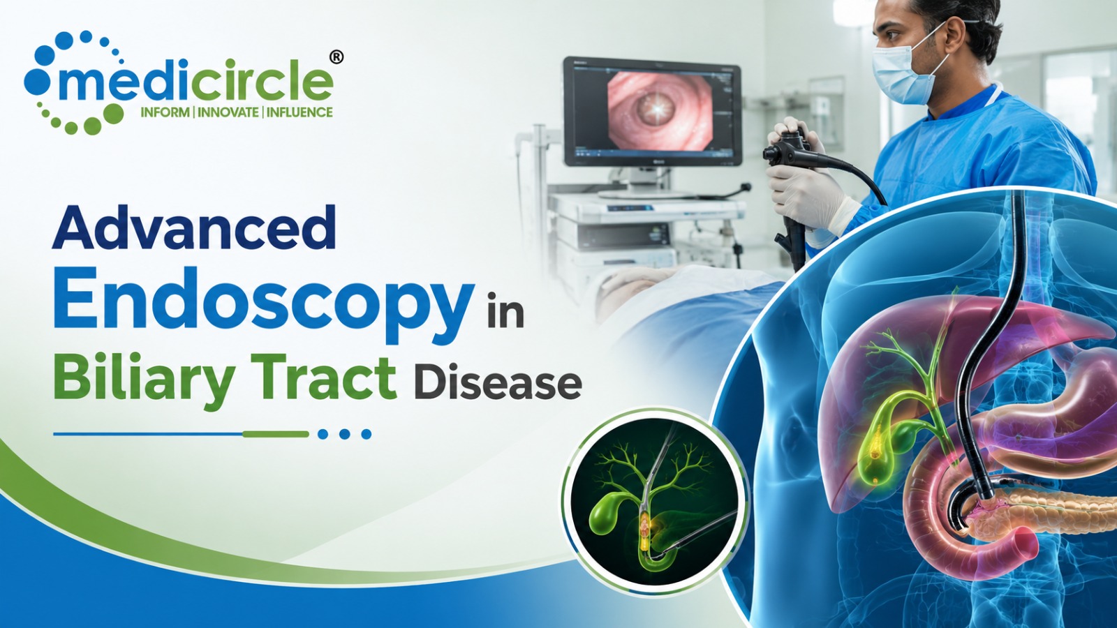
 Modern biliary disease management increasingly uses EUS and MRCP for safer diagnosis, tissue acquisition, and staging, while reserving ERCP primarily for therapeutic interventions such as stone extraction, drainage, and stenting.




Biliary tract pathologies—ranging from benign choledocholithiasis and biliary strictures to malignant cholangiocarcinomas and pancreatic head adenocarcinomas—represent major diagnostic and therapeutic challenges in gastroenterology.
Historically, Endoscopic Retrograde Cholangiopancreatography (ERCP) was utilized for both diagnosing and treating pancreatobiliary disorders. However, because diagnostic ERCP carries a documented risk of serious complications—including post-ERCP pancreatitis (PEP), cholangitis, duodenal perforation, and bleeding—the modern clinical paradigm has fundamentally shifted.
Diagnostic evaluation has largely transitioned to high-resolution, non-invasive or minimally invasive modalities: Magnetic Resonance Cholangiopancreatography (MRCP) and Diagnostic Endoscopic Ultrasound (EUS). Today, EUS serves as the premier diagnostic imaging and tissue-acquisition tool, while ERCP is reserved primarily for therapeutic decompression, stone extraction, and stent delivery.
1. Technological Foundations and Core Mechanisms
Understanding the complementary roles of EUS and ERCP requires analyzing their respective imaging and mechanical principles:
- Diagnostic Endoscopic Ultrasound (EUS):
- Mechanism: Combines a high-frequency ultrasound transducer (5–12 MHz) on the tip of a flexible endoscope with direct luminal visualization.
- Imaging Capabilities: Positioned in the duodenal bulb or stomach, EUS bypasses bowel gas and abdominal adipose tissue, providing spatial resolution (down to < 1 mm) of the common bile duct (CBD), gallbladder wall, ampulla of Vater, and regional lymph nodes.
- Tissue Acquisition: Enables real-time, Doppler-guided Fine-Needle Aspiration (FNA) or Fine-Needle Biopsy (FNB) of suspected biliary wall thickening, intraductal masses, and peri-choledochal lymphadenopathy without traversing non-target peritoneal planes.
- Endoscopic Retrograde Cholangiopancreatography (ERCP):
- Mechanism: Uses a side-viewing duodenoscope (duodenoscope elevator) to locate the major duodenal papilla, cannulate the bile duct using a hydrophilic guidewire and sphincterotome under fluoroscopy, and inject radiopaque iodinated contrast to delineate luminal architecture.
- Therapeutic Execution: Delivers targeted interventions: endoscopic sphincterotomy (ES), balloon/basket ductal clearance, mechanical lithotripsy, stricture dilation, brush cytology, and plastic or self-expanding metal stent (SEMS) deployment.
2. Choledocholithiasis: The Shift from Diagnostic Cannulation to EUS Triage
In suspected common bile duct (CBD) stones, clinical guidelines from the American Society for Gastrointestinal Endoscopy (ASGE) and European Society of Gastrointestinal Endoscopy (ESGE) categorize patients into risk strata to determine whether EUS or ERCP is the appropriate initial step.
- The Problem with Diagnostic ERCP for Stones: Diagnostic cannulation solely to confirm a stone carries a 3% to 10% risk of post-ERCP pancreatitis. Furthermore, small stones (< 4 mm) or sludge can be obscured by dense contrast medium during fluoroscopy.
- EUS Diagnostic Accuracy: EUS exhibits a sensitivity > 95% and specificity > 97% for choledocholithiasis, outperforming transabdominal ultrasound and matching or exceeding MRCP, particularly for small stones (< 5 mm) and distal duct ampullary microlithiasis.
- Same-Session EUS-to-ERCP Workflow: In intermediate-risk patients, performing an on-table EUS under the same anesthetic session allows immediate transition to therapeutic ERCP if a stone is verified, sparing approximately 60% to 70% of patients unnecessary ERCP cannulation.
3. Indeterminate Biliary Strictures: Distinguishing Benign from Malignant Disease
Evaluating focal biliary strictures requires precise tissue acquisition to differentiate cholangiocarcinoma, pancreatic adenocarcinoma, and extrinsic nodal compression from benign conditions (such as IgG4-related sclerosing cholangitis, primary sclerosing cholangitis, or post-operative ischemic strictures).
- ERCP Ductal Sampling Limitations:
- Standard ERCP utilizes trans-papillary brush cytology and intraductal forceps biopsy under fluoroscopic guidance.
- Diagnostic Sensitivity: Brush cytology has a high specificity (> 95%) but a low sensitivity (typically 30% to 50%), limited by superficial mucosal sampling, desmoplastic tumor biology, and cellular necrosis.
- EUS-Guided Tissue Acquisition (EUS-FNB):
- EUS visualizes the full extraluminal thickness of the bile duct, the surrounding vascular architecture (portal vein, hepatic artery invasion), and adjacent lymph nodes.
- Modern core-biopsy needles with reverse-bevel or Franseen geometry (EUS-FNB) obtain intact tissue architecture for immunohistochemical profiling (e.g., IgG4, CK7, CK20, Ki-67), increasing diagnostic sensitivity for malignant strictures to 85%–90%.
- Advanced Intraductal Cholangioscopy (SpyGlass):
- When EUS and standard ERCP brushing remain non-diagnostic, single-operator peroral cholangioscopy (passed through the ERCP duodenoscope directly into the biliary tree) enables direct optical visualization of the stricture mucosa and targeted "smart" biopsy of tumor vessels.
4. Structural Comparison: Operational Characteristics of EUS vs. ERCP
- Primary Clinical Purpose:
- Diagnostic EUS: High-resolution transmural visualization, staging, and tissue biopsy (FNA/FNB).
- Therapeutic ERCP: Restoring biliary drainage, stone extraction, ductal dilation, and stent placement.
- Tissue Sampling Modality:
- Diagnostic EUS: Transmural fine-needle biopsy (FNB) under real-time acoustic and Doppler guidance.
- Therapeutic ERCP: Trans-papillary brush cytology and intraductal fluoroscopy-guided forceps biopsies.
- Diagnostic Sensitivity for Malignancy:
- Diagnostic EUS: High (80% to 90% via FNB core architecture).
- Therapeutic ERCP: Moderate to low (35% to 55% via standard brushings).
- Major Adverse Event Profile:
- Diagnostic EUS: Low (< 1% risk; minor bleeding, rare perforation, near-zero pancreatitis risk).
- Therapeutic ERCP: Significant (6% to 12% total risk; post-ERCP pancreatitis, cholangitis, perforation, bleeding).
- Vascular & Nodal Staging Capability:
- Diagnostic EUS: Excellent (accurately maps tumor depth, portal vein abutment, and regional celiac/portal lymph nodes).
- Therapeutic ERCP: None (fluoroscopy visualizes intraluminal contours only).
- Direct Duct Drainage Capability:
- Diagnostic EUS: None directly (unless performing advanced therapeutic EUS-guided biliary drainage / EUS-BD).
- Therapeutic ERCP: Immediate and definitive via plastic/metal stent deployment.
5. Adverse Events and Safety Profiles: Mitigating Procedural Risk
The divergent risk profiles between EUS and ERCP dictate modern diagnostic workflows:
- Post-ERCP Pancreatitis (PEP):
- Mechanisms: Mechanical irritation of the pancreatic duct (PD) sphincter during difficult cannulation, hydrostatic injury from accidental PD contrast injection, or thermal injury during electrosurgical sphincterotomy.
- Universal Mitigation: Routine administration of rectal indomethacin or diclofenac (100 mg) immediately pre- or post-procedure, aggressive intravenous hydration with lactated Ringer's solution, and prophylactic temporary pancreatic duct stenting (5-Fr) in high-risk cannulations.
- Infectious Complications (Acute Cholangitis):
- Occurs during ERCP if contrast is injected into an obstructed, contaminated biliary system without achieving complete, unhindered drainage.
- Mandate: Avoid over-injection of contrast proximal to high-grade hilar strictures unless immediate bilateral stenting is achievable.
- Duodenal Perforation (Stapfer Classification):
- Type I (duodenal wall by endoscope tip), Type II (peri-ampullary during sphincterotomy), Type III (biliary ductal by guidewires), and Type IV (retroperitoneal air from excessive insufflation).
- EUS Safety Advantage: Diagnostic EUS avoids instrumenting the major papilla and injecting contrast under pressure, virtually eliminating the risk of pancreatitis (< 0.1%) and reducing perforation rates to < 0.05%.
6. Strategic Clinical Decision Algorithm for Biliary Tract Disease
When managing a patient presenting with obstructive jaundice, suspected choledocholithiasis, or a biliary stricture, execute a structured diagnostic sequence:
- Step 1: Clinical and Laboratory Triage: Evaluate liver function tests (total/direct bilirubin, alkaline phosphatase, ALT, AST) and assess for systemic signs of ascending cholangitis (Charcot's Triad: fever, jaundice, right upper quadrant pain).
- Step 2: Non-Invasive Cross-Sectional Imaging: Perform transabdominal ultrasound followed by multiphase contrast-enhanced CT of the abdomen/pancreas or Magnetic Resonance Cholangiopancreatography (MRCP) to delineate ductal anatomy.
- Step 3: Intermediate Choledocholithiasis Management: If MRCP is equivocal or unavailable, perform first-line EUS. If a stone is identified, proceed immediately to same-session therapeutic ERCP with sphincterotomy and balloon extraction. If negative, avoid ERCP entirely.
- Step 4: Indeterminate Stricture Evaluation & Staging: Perform Diagnostic EUS with FNB as the initial invasive test to assess parenchymal masses, vascular invasion, and lymph nodes, obtaining a tissue core for pathology.
- Step 5: Therapeutic Decompression: Once tissue diagnosis is secured or if the patient presents with cholangitis/severe pruritus, perform Therapeutic ERCP for stent placement (fully covered SEMS for benign strictures; uncovered/covered SEMS or plastic stents for unresectable malignancy).
- Step 6: Refractory Access (EUS-Guided Biliary Drainage): If ERCP cannulation fails due to altered surgical anatomy (e.g., Roux-en-Y gastric bypass, Whipple resection) or severe duodenal tumor infiltration, perform advanced EUS-guided Biliary Drainage (EUS-BD) via hepaticogastrostomy or choledochoduodenostomy using lumen-apposing metal stents (LAMS).
10 Frequently Asked Questions (FAQs)
Q1. Why is diagnostic ERCP no longer standard clinical practice?Diagnostic ERCP has been largely replaced because it carries a substantial 3% to 10% risk of acute post-ERCP pancreatitis, along with risks of infection, hemorrhage, and perforation. Non-invasive MRCP and low-risk diagnostic EUS provide superior or equivalent imaging detail without these complications.
Q2. How does rectal indomethacin prevent post-ERCP pancreatitis (PEP)?Rectal indomethacin (100 mg administered per rectum at the time of the procedure) is a potent non-steroidal anti-inflammatory drug that inhibits phospholipase A2 and cyclooxygenase (COX) pathways, reducing the early inflammatory cascade responsible for acute post-cannulation pancreatitis.
Q3. What is the diagnostic accuracy of EUS for detecting small common bile duct stones?EUS has a sensitivity of 95% to 98% and a specificity of 97% to 100% for choledocholithiasis, demonstrating particular superiority over transabdominal ultrasound and CT for detecting microlithiasis and small stones less than 5 mm located in the distal common bile duct.
Q4. What is the difference between EUS-FNA and EUS-FNB?EUS-FNA (Fine-Needle Aspiration) uses standard hollow-bore needles to collect individual cells for cytology. EUS-FNB (Fine-Needle Biopsy) utilizes specialized needle tips (e.g., Fork-tip, Franseen) to core out intact histologic tissue specimens, preserving cellular architecture and enabling comprehensive immunohistochemical and molecular analysis.
Q5. When is MRCP preferred over EUS as the initial diagnostic test?MRCP is completely non-invasive, requires no sedation, and provides a global overview of the entire intrahepatic and extrahepatic biliary tree. It is preferred as the initial non-invasive test for broad anatomical mapping or when patients have contraindications to conscious sedation or upper endoscopy.
Q6. What is SpyGlass cholangioscopy, and when is it indicated?SpyGlass is a single-operator peroral cholangioscope passed through the working channel of an ERCP duodenoscope directly into the bile ducts. It is indicated for direct optical visualization and targeted biopsy of indeterminate strictures, as well as electrohydraulic or laser lithotripsy of large, impacted bile duct stones.
Q7. What are the signs of post-ERCP pancreatitis (PEP)?PEP is characterized by new-onset, severe, persistent epigastric pain radiating to the back, accompanied by a serum amylase or lipase elevation greater than 3 times the upper limit of normal measured 24 hours post-procedure, often requiring prolonged hospitalization.
Q8. What is EUS-guided Biliary Drainage (EUS-BD)?EUS-BD is an advanced rescue technique used when conventional ERCP cannulation fails. Under combined endoscopic ultrasound and fluoroscopic guidance, the biliary tree is punctured from the stomach (hepaticogastrostomy) or duodenum (choledochoduodenostomy), and a stent is deployed across the tract to restore biliary outflow.
Q9. How do clinicians manage malignant biliary obstruction at the liver hilum (Klatskin tumor)?Malignant hilar obstructions require careful pre-procedural planning using high-resolution CT/MRCP to map which hepatic segments need drainage. Drainage is executed using specialized unilateral or bilateral plastic/metal stenting during ERCP, taking care to avoid injecting contrast into non-drained hepatic segments to prevent fatal cholangitis.
Q10. What is a "difficult cannulation" during ERCP, and what are the rescue options?Difficult cannulation is defined by guidelines as more than 5 minutes of attempted cannulation, more than 5 attempts, or more than 1 to 2 unintended pancreatic duct passages. Rescue options include using a double-guidewire technique, needle-knife precut papillotomy, transpancreatic septotomy, or transitioning to EUS-guided rendezvous techniques.






.jpeg)









.jpg)
.jpg)
.jpg)
.jpg)
.jpg)
.jpg)


.jpg)