 Friday, October 09, 2026
 India’s four New Labour Codes reshape wages, social security, working hours, industrial relations, and workplace compliance, requiring employers to modernize payroll, HR systems, contracts, attendance, and employee-management processes.




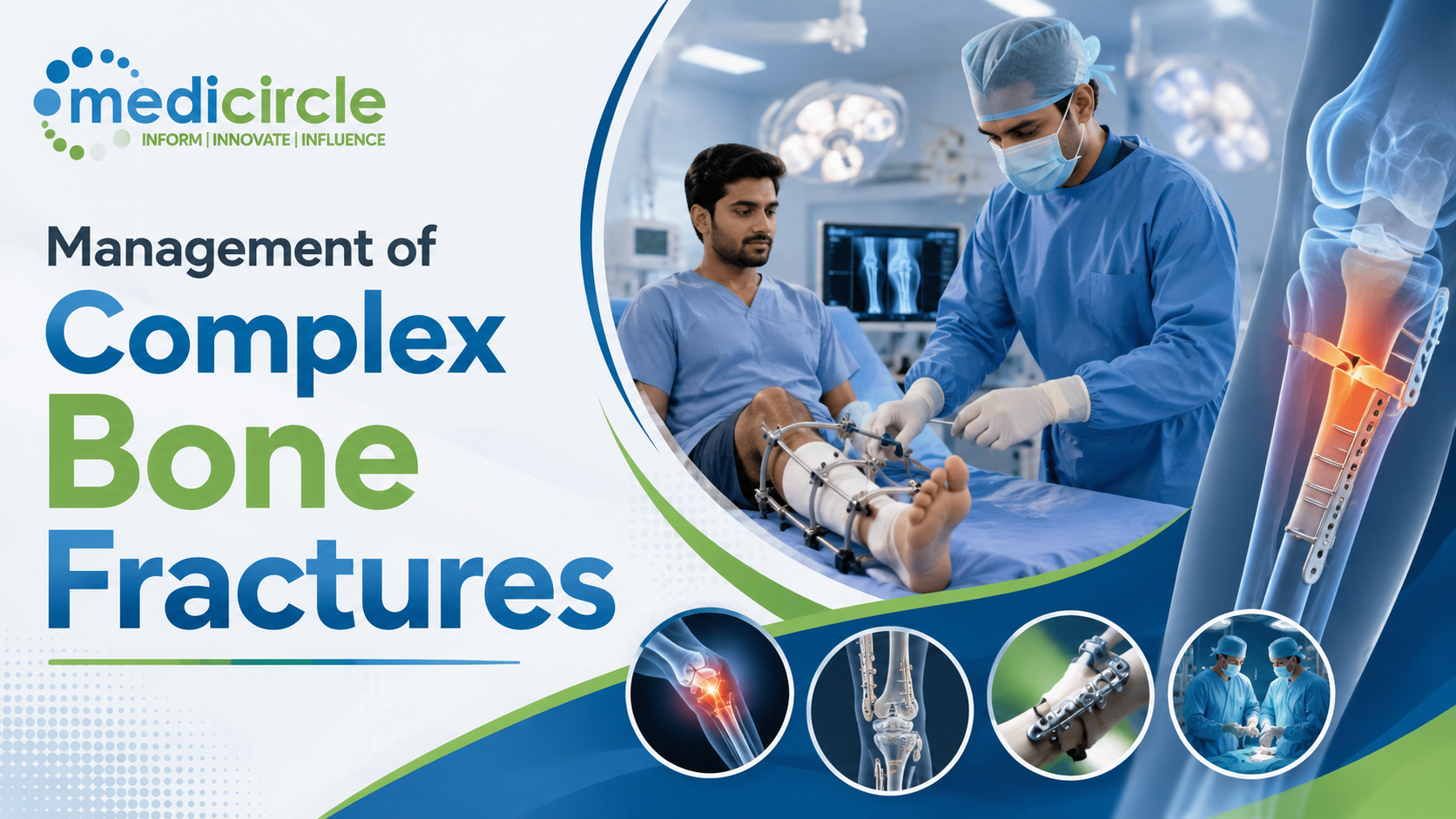
Complex skeletal fractures—characterized by high-energy comminution, severe periosteal stripping, extensive soft-tissue envelopes compromise (Gustilo-Anderson high-grade open fractures), segmental bone loss, and intra-articular disruption—represent major surgical challenges in orthopedic and trauma reconstructive surgery.
When mechanical stability or biological vascularity is compromised, bone healing stalls. According to the FDA and international orthopedic definitions, a delayed union occurs when a fracture fails to consolidate within the expected timeframe (typically 3 to 6 months), while a non-union is established when at least 9 months have elapsed since the injury with no visible radiological signs of progressive healing over 3 consecutive months.
Resolving complex non-unions and critical-sized segmental bone defects requires a dual biomechanical and biological strategy: applying the Diamond Concept of bone healing, selecting autologous vs. synthetic bone graft matrices, and implementing modern fixation innovations (such as anatomically pre-contoured locking compression plates, suprapatellar intramedullary nails, and the Masquelet induced-membrane technique).
1. Pathophysiology and Classification of Fracture Non-Unions
Fracture consolidation depends on the cellular interplay between mechanical strain and local biological vascularity (Perren's Strain Theory). Understanding why a fracture fails to unite dictates the surgical corrective approach.
- Hypertrophic Non-Union ("Elephant's Foot" / "Horse's Hoof"):
- Biological State: Excellent biology and rich local vascularity, but inadequate mechanical stability (excessive interfragmentary motion/strain > 10% to 15%).
- Radiological Features: Abundant, flared, unbridged callus formation with a persistent radiolucent fracture line and viable, sclerotic bone ends.
- Corrective Principle: Requires strictly mechanical revision—rigid internal or external fixation to eliminate micromotion; biological bone grafting is typically unnecessary.
- Atrophic Non-Union ("Pencil-in-Cup" / Inert Bone Ends):
- Biological State: Avascular, biologically inert fracture gap resulting from extensive periosteal stripping, thermal bone necrosis, severe soft-tissue degloving, or microvascular compromise (e.g., in smokers, uncontrolled diabetics, or post-radiation bone).
- Radiological Features: Complete absence of callus formation, tapered/osteopenic bone ends, rounded bone margins, and a wide fibrous gap.
- Corrective Principle: Requires complete decortication of necrotic bone ends, opening the sclerotic medullary canals, and delivering rich biological bone grafts alongside rigid internal fixation.
- Oligotrophic Non-Union:
- Biological State: Intermediate state with minimal callus due to poor mechanical apposition (e.g., large distraction gap following initial traction or plate fixation) despite intact vascularity.
- Infected / Septic Non-Union:
- Pathology: Bacterial biofilm formation (predominantly Staphylococcus aureus or Pseudomonas aeruginosa) across necrotic cortical bone (sequestrum) and hardware implants.
- Mandate: Requires a staged radical debridement, implant removal, dead space management with antibiotic cement spacers, pathogen-directed systemic antimicrobial therapy, and delayed reconstruction.
2. The Diamond Concept of Bone Healing and Grafting Strategies
The modern standard for skeletal tissue regeneration is the Diamond Concept (formulated by Giannoudis et al.), which establishes that successful bone consolidation requires five interconnected elements:
- 1. Osteogenic Cells: Viable progenitor cells capable of differentiating into osteoblasts (e.g., skeletal stem cells from bone marrow aspirate).
- 2. Osteoinductive Mediators: Growth factors that recruit and stimulate stem cell differentiation into osteoblasts (e.g., Recombinant Human Bone Morphogenetic Proteins: rhBMP-2, rhBMP-7, Platelet-Derived Growth Factor).
- 3. Osteoconductive Matrix / Scaffold: A three-dimensional physical scaffold that supports the ingrowth of newly forming capillaries and bone trabeculae (e.g., mineralized extracellular matrix, hydroxyapatite, beta-tricalcium phosphate).
- 4. Mechanical Stability: Rigid or dynamic fixation that maintains interfragmentary strain within the optimal physiological window (2% to 10%) for primary or secondary bone healing.
- 5. Vascularity / Host Bed Vascular Environment: Healthy surrounding soft tissue and muscle envelopes providing blood supply and local oxygen tension.
3. Structural Comparison: Bone Grafting Materials and Biological Matrices
- Autologous Cancellous Bone Graft (Iliac Crest Bone Graft / ICBG):
- Osteogenic Potential: Present (contains live osteoprogenitor cells).
- Osteoinductive Potential: Present (contains native BMPs, TGF-beta, and VEGF).
- Osteoconductive Potential: Present (natural porous trabecular scaffold).
- Mechanical Load Support: Negligible / Low.
- Primary Limitation: Donor-site morbidity (chronic pain, hematoma, nerve paresthesia) and limited harvest volume.
- Clinical Utility: Gold standard for bridging non-unions and small-to-moderate cavitary defects (< 3 to 5 cm).
- Reamer-Irrigator-Aspirator (RIA) Autograft (Femoral/Tibial Canal):
- Osteogenic Potential: Very High (high concentration of mesenchymal stem cells and endosteal marrow).
- Osteoinductive Potential: High (rich in growth factors).
- Osteoconductive Potential: Present (morcellized cortical/cancellous debris).
- Mechanical Load Support: None.
- Primary Limitation: Requires dedicated intramedullary reaming hardware; risk of cortical perforation or fat embolism.
- Clinical Utility: Preferred source for large-volume (> 30–50 mL) autologous grafting in complex non-unions.
- Allograft (Fresh-Frozen or Freeze-Dried Cancellous/Cortical Bone):
- Osteogenic Potential: Absent (cells devitalized during sterilization).
- Osteoinductive Potential: Minimal to low.
- Osteoconductive Potential: Excellent (intact human mineral lattice).
- Mechanical Load Support: Moderate to High (structural cortical allograft struts).
- Primary Limitation: Slower incorporation, risk of immune rejection or rare disease transmission.
- Clinical Utility: Structural reconstruction of large diaphyseal/metaphyseal defects and impaction grafting.
- Demineralized Bone Matrix (DBM):
- Osteogenic Potential: Absent.
- Osteoinductive Potential: Moderate (acid extraction exposes embedded collagen-bound BMPs).
- Osteoconductive Potential: Moderate.
- Mechanical Load Support: None (putty, gel, or paste consistency).
- Primary Limitation: High batch-to-batch variability in growth factor potency.
- Clinical Utility: Bone graft extender paired with autologous bone aspirate.
- Synthetic Ceramic Substitutes (Hydroxyapatite / Beta-TCP / Bioglass):
- Osteogenic Potential: Absent.
- Osteoinductive Potential: Absent (unless bio-engineered with recombinant growth factors).
- Osteoconductive Potential: Excellent (calcium phosphate mineral lattice).
- Mechanical Load Support: Good in compression, brittle in shear/torsion.
- Primary Limitation: Slow or incomplete resorption rates; risks foreign-body reaction if exposed.
- Clinical Utility: Filling benign cystic cavities or extending autologous harvests.
- Recombinant Human Bone Morphogenetic Protein-2 (rhBMP-2 / Infuse):
- Osteogenic Potential: Indirect (actively recruits surrounding mesenchymal cells).
- Osteoinductive Potential: Exceptionally High.
- Osteoconductive Potential: Supplied with an absorbable collagen sponge (ACS) carrier.
- Mechanical Load Support: None.
- Primary Limitation: High cost; risk of heterotopic ossification, severe localized seroma/edema.
- Clinical Utility: Acute open tibial shaft fractures and refractory recalcitrant non-unions.
4. Fixation Innovations: Biomechanics of Locking Plates, Nails, and Induced Membranes
Modern trauma surgery relies on biomechanically advanced hardware configurations to stabilize difficult fracture patterns without compromising periosteal microvascular perfusion:
- Locking Compression Plates (LCP) and Far Cortical Locking:
- Internal Fixator Principle: Screws thread directly into the plate holes, creating a fixed-angle construct. The plate does not need to be compressed tightly against the bone surface, preserving the underlying periosteal capillary network.
- Far Cortical Locking (FCL): Screws engage only the opposite (far) cortex while passing unthreaded through the near cortex. This allows controlled, parallel micro-motion across both cortices, preventing asymmetric stiffening and promoting circumferential secondary callus formation.
- Suprapatellar Intramedullary Nailing (Semi-Extended Technique):
- Insertion of tibial intramedullary nails via a suprapatellar approach with the knee in semi-extension (15 to 20 degrees flexion).
- Biomechanical Advantage: Eliminates the quadriceps deforming forces that cause apex-anterior (procurvatum) and valgus malalignment in proximal-third and distal-third tibial fractures, simplifying fracture reduction and fluoroscopic imaging.
- The Masquelet Induced-Membrane Technique (For Critical-Sized Defects > 4–5 cm):
- Stage 1: Radical debridement of all devitalized bone; placement of a polymethylmethacrylate (PMMA) antibiotic-loaded bone cement spacer across the defect, followed by rigid fixation. Over 6 to 8 weeks, the body generates a vascularized, biological "pseudo-synovial" foreign-body membrane around the cement block.
- Stage 2: Incision of the membrane, removal of the cement spacer without damaging the envelope, packing the cavity with high-density autologous bone graft (RIA/ICBG), and closing the biological membrane. The induced membrane secretes VEGF, TGF-beta, and BMP-2 while protecting the graft from resorption, enabling rapid consolidation of defects up to 10 to 15 cm.
5. Strategic Step-by-Step Surgical Decision Protocol
When evaluating a patient with a complex fracture non-union or segmental bone defect, execute this five-stage surgical management sequence:
- Step 1: Diagnostic Workup and Infection Exclusion: Obtain orthogonal plain radiographs and high-resolution thin-cut CT with 3D reconstructions. Rule out occult infection by evaluating serum ESR, CRP, and performing image-guided aspiration for microbiology if inflammatory markers are elevated.
- Step 2: Optimize Host Systemic Factors: Enforce strict tobacco/nicotine cessation (nicotine impairs microvascular perfusion and osteoblast function); optimize glycemic control (HbA1c < 7.0\%), and correct 25-hydroxyvitamin D (> 30\text{ ng/mL}) and calcium levels.
- Step 3: Surgical Exposure and Radical Biologic Debridement: Excise all dense avascular scar tissue; resect necrotic cortical bone back to bleeding margins ("Paprika sign"); recanalize sclerotic intramedullary canals with sequential drills/curettes until endosteal vascular flow is restored.
- Step 4: Execute Targeted Reconstruction:
- For pure hypertrophic non-union: Revise hardware to a high-rigidity construct (e.g., exchanged reamed intramedullary nail with larger diameter or locked compression plating).
- For atrophic non-union with minimal bone loss (< 2 cm): Stable locked plating paired with local autologous cancellous bone grafting (RIA or ICBG).
- For critical-sized segmental bone loss (> 4–5 cm): Initiate the 2-stage Masquelet Induced-Membrane Protocol or vascularized free fibular transfer.
- Step 5: Post-Operative Biomechanical Progression: Initiate early, progressive protected weight-bearing based on construct mechanics, using periodic CT assessments at 12 and 24 weeks to confirm trabecular bridging before full unassisted loading.
10 Frequently Asked Questions (FAQs)
Q1. What is the clinical definition of a fracture non-union?A non-union is formally diagnosed when a fractured bone fails to achieve bony consolidation after a minimum of 9 months from the initial injury, provided that serial radiographs show no progressive signs of healing over a consecutive 3-month observation window.
Q2. How do clinicians differentiate between a hypertrophic and an atrophic non-union?Hypertrophic non-unions display abundant, flared callus formation ("elephant's foot") on radiographs, indicating healthy biological vascularity that is failing solely due to mechanical instability. Atrophic non-unions display absent callus, tapered bone ends, and poor local blood supply, requiring biological bone grafting in addition to mechanical stabilization.
Q3. Why is autologous bone graft considered superior to synthetic ceramic bone substitutes?Autologous bone graft (from the iliac crest or intramedullary canal) is the only grafting material that contains all three primary regenerative properties: osteogenesis (live bone-forming cells), osteoinduction (native growth factors like BMPs), and osteoconduction (natural trabecular matrix). Synthetic substitutes provide only osteoconduction.
Q4. What is the Reamer-Irrigator-Aspirator (RIA) system?RIA is a specialized surgical device used inside the intramedullary canal of long bones (typically the femur). It continuous irrigates and aspirates while reaming the canal, harvesting large volumes (30 to 60 mL) of high-potency autologous bone graft and mesenchymal stem cells while minimizing thermal necrosis and fat embolism risk.
Q5. How does smoking/nicotine use affect fracture healing and non-union rates?Nicotine is a potent peripheral vasoconstrictor that diminishes microvascular blood flow to the periosteum and fracture callus. It also inhibits osteoblast proliferation, increases platelet aggregation, and downregulates collagen synthesis, increasing non-union rates by two- to three-fold.
Q6. What is the Masquelet technique, and when is it indicated?The Masquelet technique is a two-stage surgical procedure used to reconstruct large segmental bone defects (> 4 to 5 cm). Stage 1 involves debridement and placement of an antibiotic PMMA cement spacer, which induces a biological, vascularized membrane. Stage 2 (6 to 8 weeks later) replaces the cement with autologous bone graft inside the vascular envelope.
Q7. What role does Vitamin D play in complex fracture healing?Vitamin D (specifically its active form, 1,25-dihydroxyvitamin D) is required for intestinal calcium absorption and mineralized bone matrix deposition. Serum 25-hydroxyvitamin D levels below 30 ng/mL are associated with delayed callus mineralization, reduced mechanical torsional strength of healing bone, and increased non-union rates.
Q8. What is the difference between primary (direct) and secondary (indirect) bone healing?- Primary (Direct) Healing: Occurs with anatomical reduction and absolute stability (rigid compression plating, strain < 2%). Healing proceeds via direct osteonal cutting cones across the fracture line without external callus.
- Secondary (Indirect) Healing: Occurs with relative stability (intramedullary nailing, external fixation, bridging plates, strain 2% to 10%). Healing proceeds through endochondral ossification with visible, robust periosteal callus formation.
Traditional non-locking plates rely on friction, requiring the plate to be compressed tightly against the bone surface by standard screws, which crushes the underlying periosteal capillary network. Locking plates lock the screw heads into the plate holes, functioning as an "internal fixator" elevated slightly above the cortex, preserving periosteal blood flow.
Q10. What is a "sequestrum" in an infected non-union?A sequestrum is a fragment of devitalized, necrotic cortical bone that has separated from healthy vascularized bone during chronic osteomyelitis. Because it lacks a blood supply, systemic antibiotics cannot reach the bacteria embedded within its biofilm, requiring complete surgical debridement (sequestrectomy) before bone healing can proceed.






.jpeg)







.jpg)
.jpg)
.jpg)
.jpg)
.jpg)
.jpg)

.jpg)