Tuesday, October 06, 2026
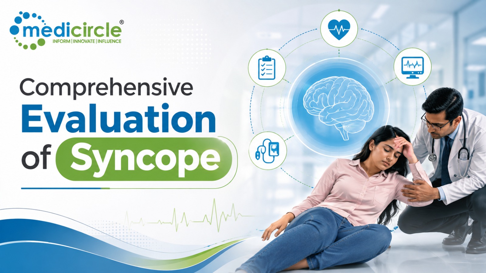
 Syncope—defined as a transient loss of consciousness (TLOC) due to global cerebral hypoperfusion, characterized by rapid onset, short duration, and spontaneous complete recovery—presents a major diagnostic challenge in emergency and outpatient clinical medicine.




Syncope—defined as a transient loss of consciousness (TLOC) due to global cerebral hypoperfusion, characterized by rapid onset, short duration, and spontaneous complete recovery—presents a major diagnostic challenge in emergency and outpatient clinical medicine.
The primary clinical imperative during syncope evaluation is risk stratification: distinguishing benign reflex mechanisms from life-threatening cardiac etiologies or non-syncopal neurological mimics (such as epilepsy).
1. Pathophysiological Classification & Etiology
Syncope is broadly categorized into three true pathophysiological mechanisms, alongside non-syncopal mimics:
A. Reflex (Neurally Mediated) SyncopeThe most common cause across all age groups. It results from inappropriate autonomic reflex responses triggering vasodilation (vasodepressor response) and/or bradycardia (cardioinhibitory response).
- Vasovagal Syncope (VVS): Triggered by prolonged standing, heat, or emotional stress/pain.
- Situational Syncope: Triggered by specific physiological acts (micturition, deglutition, coughing, defecation).
- Carotid Sinus Syndrome: Hypersensitivity of carotid baroreceptors triggered by neck turning or tight collars.
Results from a sudden, critical drop in cardiac output due to electrical or structural abnormalities. Associated with high 1-year mortality and sudden cardiac death (SCD).
- Arrhythmias (Electrical): Bradyarrhythmias (Sick Sinus Syndrome, High-grade AV block) or Tachyarrhythmias (Ventricular Tachycardia, Brugada Syndrome, Long QT Syndrome, Wolff-Parkinson-White Syndrome).
- Structural Heart Disease: Aortic Stenosis, Hypertrophic Cardiomyopathy (HCM), Acute Myocardial Infarction, Pulmonary Embolism, Aortic Dissection, or Cardiac Tamponade.
Occurs when systemic vasoconstrictive reflexes fail upon standing, resulting in an uncompensated venous pooling.
- Causes: Volume depletion (dehydration, hemorrhage), primary autonomic failure (Parkinson’s disease, Multiple System Atrophy), or secondary autonomic failure (diabetic neuropathy, vasodilator/diuretic medications).
Conditions that present with loss of consciousness or pseudo-unconsciousness but are not caused by transient global cerebral hypoperfusion.
- Epileptic Seizures: Primary or secondary electrical hypersynchrony.
- Vertebrobasilar Transient Ischemic Attack (TIA): Rare cause of isolated TLOC; usually accompanied by focal neurological deficits (diplopia, dysarthria, ataxia).
- Psychogenic Pseudosyncope (PPS): Conversion disorder or malingering; normal EEG and vitals during episodes.
2. Diagnostic Differential: Syncope vs. Seizure vs. Cardiac Arrest
Differentiating true syncopal events from neurological or electrical conditions is the first step in clinical evaluation:
- Prodromal Symptoms: Reflex syncope features progressive lightheadedness, nausea, diaphoresis, tunnel vision, and pallor. Cardiac syncope often presents with brief or absent prodrome (sudden "drop attack") or sudden palpitations. Seizures feature stereotyped motor/sensory auras (e.g., epigastric rising, olfactory hallucinations).
- Onset Dynamics: Reflex and OH syncope occur during upright posture or specific triggers. Cardiac syncope can occur during exertion or while supine. Seizures occur in any posture or during sleep.
- Episode Duration: Syncope lasts seconds to less than 1–2 minutes. Seizures typically last 1 to 3 minutes or longer.
- Post-Event Recovery: Syncope returns rapidly to baseline orientation without persistent confusion. Seizures feature a prolonged post-ictal state (confusion, fatigue, amnesia, focal weakness/Todd's paralysis).
- Associated Signs: Muscle tone is flaccid in syncope (though brief myoclonic jerks can occur during prolonged cerebral hypoxia). Seizures feature sustained tonic-clonic motor activity, lateral tongue biting, and urinary incontinence.
3. The High-Yield Diagnostic Workup Pipeline
A structured, risk-stratified diagnostic pathway prevents unnecessary neuroimaging while ensuring rapid cardiac intervention for high-risk patients.
Step 1: Initial Emergency Triage & Risk Stratification- Detailed History & Bystander Accounts: Establish posture, triggers, prodrome, duration, and recovery phase.
- Physical Exam & Orthostatic Vitals: Measure blood pressure and heart rate in supine position and after 1 and 3 minutes of standing (positive OH: drop in \text{SBP} \ge 20\text{ mmHg} or \text{DBP} \ge 10\text{ mmHg}).
- 12-Lead Electrocardiogram (ECG): Mandatory for 100% of syncope patients. Screen for PR interval, QRS duration, QTc prolongation/shortening, Brugada pattern (V_1\text{--}V_2 ST elevation), delta waves (WPW), Q waves, and arrhythmogenic right ventricular cardiomyopathy (ARVC) epsilon waves.
Indicated if history, physical exam, or ECG suggests cardiac origin, or if high-risk red flags are present (exertional syncope, supine syncope, family history of sudden cardiac death, or structural heart disease):
- Echocardiography: Assess for valvular lesions (Aortic Stenosis), left ventricular ejection fraction (LVEF), wall motion abnormalities, or HCM.
- Ambulatory ECG Monitoring:
- Holter Monitor (24–48 hours): For frequent daily symptoms.
- Event Recorder / Patch Monitor (14–30 days): For weekly/monthly symptoms.
- Invasive Implantable Loop Recorder (ILR): The gold standard for recurrent, unexplained, high-risk syncope with low baseline frequency.
- Invasive Electrophysiology Study (EPS): Indicated in patients with previous MI, structural heart disease, or unexplainable bundle branch block to evaluate sinus node recovery time and induce ventricular arrhythmias.
Indicated only when focal neurological signs are present, post-ictal states are documented, or non-syncopal TLOC (epilepsy) is suspected.
- Electroencephalogram (EEG): Routine EEG or long-term video-EEG monitoring to capture epileptiform discharges.
- Neuroimaging (Brain MRI / CT): Indicated for focal neurological deficits or acute head trauma secondary to a fall. Routine brain CT/MRI is inappropriate for isolated syncope.
- Carotid & Vertebrobasilar Duplex Ultrasound: Indicated only if focal brainstem signs (dizziness, dysarthria, diplopia) accompany the event.
- Head-Up Tilt-Table Testing (HUTT): Indicated to confirm vasovagal syncope, delayed orthostatic hypotension, or psychogenic pseudosyncope in diagnostic dilemmas.
- Carotid Sinus Massage (CSM): Conducted under continuous ECG and BP monitoring in patients >40 years with unexplained syncope (positive if ventricular asystole >3\text{ seconds} or SBP drop >50\text{ mmHg}).
4. Operational Action Plan for Emergency & Outpatient Teams
To optimize diagnostic yield and avoid unnecessary expenditure on low-yield tests, clinical teams should follow a three-phase operational pathway:
- Execute History, Orthostatic Vitals, and 12-Lead ECG
Phase 1: Initial Triage & TLOC Differentiation
Obtain detailed bystander history, perform supine/standing orthostatic vitals, and analyze a 12-lead ECG. Screen for high-risk red flags (exertional onset, supine syncope, structural heart disease, abnormal ECG). - Escalate to Cardiac vs. Autonomic / Neuro Diagnostics
Phase 2: Risk-Stratified Pathway Selection
If red flags or cardiac findings are present, admit to telemetry or order echocardiography and ambulatory ECG monitoring (Patch/ILR). If vasovagal features predominate with a normal ECG, reassure the patient or order Head-Up Tilt-Table Testing (HUTT). - Reserve EEG and Neuroimaging for Suspected Seizures or TIAs
Phase 3: Targeted Neurological Escalation
Order video-EEG or brain MRI only if the event features sustained tonic-clonic convulsions, prolonged post-ictal confusion, localized aura, or persistent focal neurological deficits.
Actionable Strategy: Digital Governance & Health Record Tracking
- Sync Diagnostic Records via Universal Digital Health ID Pipelines: Ensure baseline 12-lead ECGs, echocardiogram reports, and ambulatory telemetry logs are uploaded directly to national digital health accounts—such as the ABHA ID (Ayushman Bharat Health Account) network. This guarantees that longitudinal cardiac telemetry remains accessible during emergency admissions.
- Verify Specialist Credentials via Central Academic Repositories: Ensure attending electrophysiologists, neurologists, and emergency physicians maintain verified board certifications and advanced life support qualifications logged through national education registries like the APAAR ID system within the Academic Bank of Credits (ABC) network.
- Establish Rapid Telemetry Audit Protocols: Emergency departments should maintain an automated "Syncope Clinical Decision Pathway," auditing adherence to ESC/AHA guidelines to prevent low-yield routine brain CT scans in simple vasovagal cases.
Frequently Asked Questions (FAQs)
Q1. What is the single most important diagnostic test in evaluating syncope?The 12-lead Electrocardiogram (ECG) is the single most essential initial diagnostic test. It is low-cost, immediate, and can uncover life-threatening cardiac conditions such as Long QT Syndrome, Brugada Syndrome, high-grade AV block, or acute ischemia.
Q2. Is a routine head CT scan recommended for evaluating syncope?No. Routine head CT scans have an extremely low diagnostic yield (<2\%) in isolated syncope and contribute to unnecessary radiation and costs. Neuroimaging is indicated only if the patient has focal neurological deficits, head trauma from a fall, or clear seizure characteristics.
Q3. What is an Implantable Loop Recorder (ILR), and when should it be used?An ILR is a subcutaneous, long-term ECG monitoring device that records heart rhythm continuously for up to 3 years. It is recommended for patients with recurrent, unexplained syncope who lack immediate high-risk criteria requiring hospital admission, or in whom cardiac arrhythmias are strongly suspected but infrequent.
Q4. Can convulsive jerks occur during a simple vasovagal faint?Yes. Short, unsynchronized myoclonic jerks (known as "convulsive syncope") can occur during any syncopal event if cerebral perfusion is briefly deprived. Unlike an epileptic seizure, convulsive syncope lasts less than 15 seconds, lacks a prolonged post-ictal state, and features rapid recovery.
Q5. What defines a positive Head-Up Tilt-Table Test (HUTT)?A positive HUTT reproduces the patient's typical syncope accompanied by severe hypotension (vasodepressor response), bradycardia or asystole (cardioinhibitory response), or both (mixed response).
Q6. How does an ABHA ID assist patients with recurrent syncope?An ABHA ID digitizes a patient's medical history, making prior ECGs, echocardiograms, and loop recorder tracking data instantly accessible to emergency physicians across different healthcare centers.
Q7. What are the key "red flag" features suggesting cardiac syncope?Red flags include syncope occurring during physical exertion, syncope occurring in a supine position, sudden onset without prodrome, preceding palpitations, known history of structural heart disease, or a family history of unexplained sudden cardiac death at a young age.
Q8. What is Psychogenic Pseudosyncope (PPS)?Psychogenic Pseudosyncope is a non-epileptic, non-syncopal disorder where patients experience apparent loss of consciousness without underlying cerebral hypoperfusion or epileptiform brain activity. It is diagnosed using video-EEG showing normal alpha rhythm during clinical episodes.
Q9. What is Carotid Sinus Massage, and who should receive it?Carotid Sinus Massage involves manual pressure over the carotid artery bifurcation for 5 seconds to evaluate for carotid sinus hypersensitivity. It is recommended for patients over 40 years old with unexplained syncope, but is contraindicated in patients with carotid bruits or recent stroke/TIA.
Q10. What immediate action should be taken if a patient faints in front of you?Lay the patient flat on their back (supine) and elevate their legs to promote venous return to the heart. Loosen tight clothing around the neck, ensure an open airway, and monitor for normal breathing and pulse. Avoid sitting them up prematurely.






.jpeg)
.jpg)
.jpg)
.jpg)
.jpg)
.jpg)
.jpg)


.jpg)
.jpg)
.jpg)
.jpg)
.jpg)
.jpg)