 Thursday, November 05, 2026
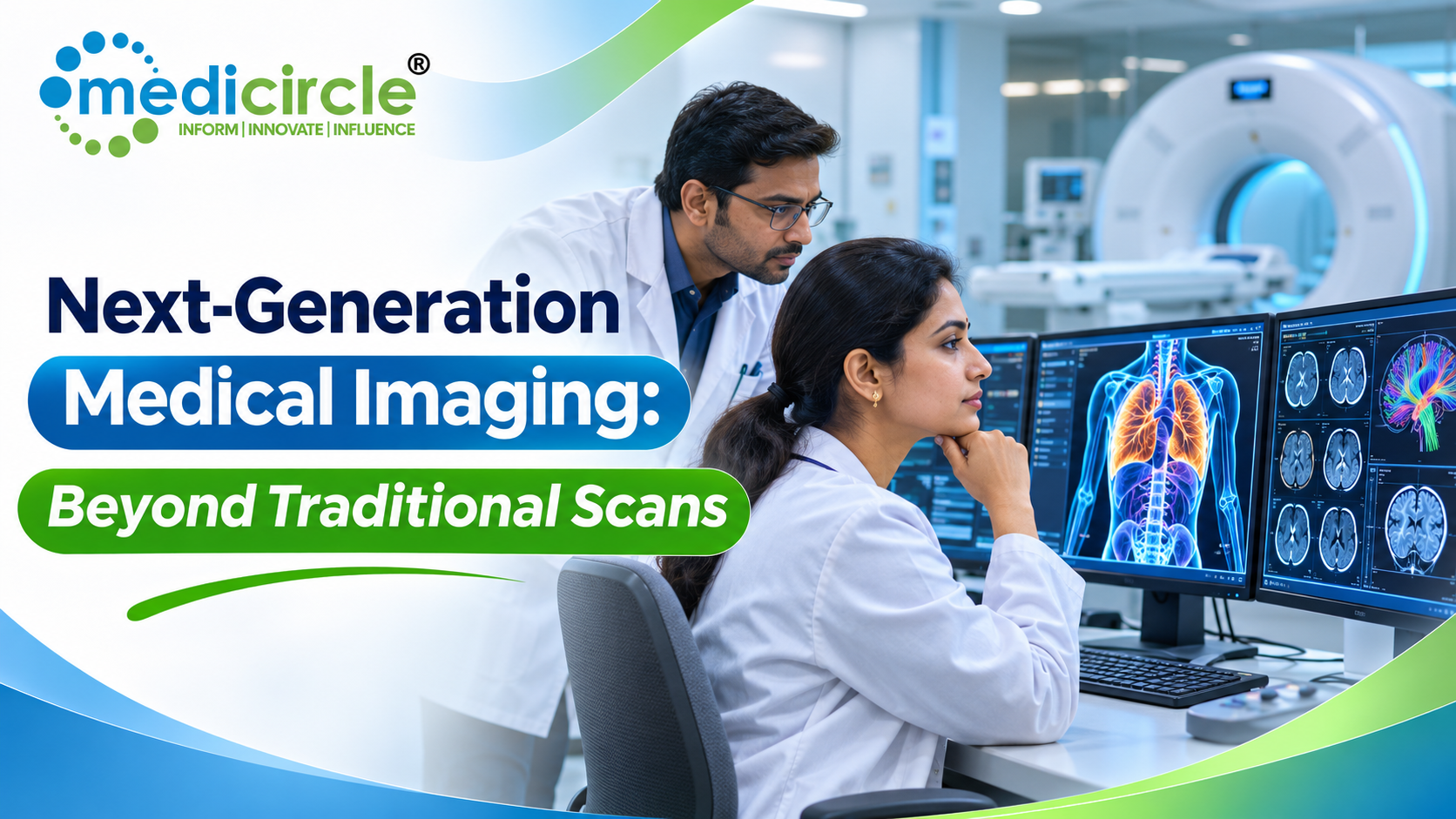
 Next-generation medical imaging technologies, including AI-powered diagnostics, molecular imaging, and portable tools, are transforming disease detection and reshaping healthcare delivery across India.





Next-Generation Imaging: What Comes After Traditional Medical Scans?
Introduction
Medical imaging has long been one of the most powerful tools in the hands of clinicians. From the first X-ray captured by Wilhelm Roentgen in 1895 to the sophisticated MRI and CT scanners that fill modern hospitals today, imaging technology has continuously evolved to give doctors a clearer, more detailed view of what is happening inside the human body.
Yet the pace of change in recent years has been extraordinary. The limitations of conventional imaging, including radiation exposure, limited soft tissue resolution, inability to detect disease at a cellular level, and long scan times, have pushed researchers, engineers, and clinicians to look beyond what traditional scans can offer.
India, with its rapidly growing healthcare infrastructure, rising burden of chronic disease, and a government actively investing in healthtech under flagship programmes such as Ayushman Bharat and the Ayushman Bharat Digital Mission (ABDM), stands at a pivotal moment in this imaging revolution. Understanding what comes after traditional medical scans is no longer a concern for radiologists alone. It is a conversation that touches every corner of the healthcare ecosystem, from patients in metro hospitals to doctors in district health centres.
Understanding Traditional Medical Imaging and Its Limitations
To appreciate what comes next, it is important to understand where the current generation of imaging stands and where it falls short.
Conventional imaging modalities include:
- X-rays: useful for bone fractures and chest evaluations but offer limited soft tissue detail and involve ionising radiation.
- Computed Tomography (CT) scans: faster and more detailed than X-rays but associated with significantly higher radiation doses.
- Magnetic Resonance Imaging (MRI): excellent for soft tissue, no radiation, but expensive, time-consuming, and unsuitable for patients with certain metallic implants.
- Ultrasound: safe and widely accessible but operator-dependent and limited in depth resolution.
- Nuclear medicine scans such as PET: highly functional but costly and limited in availability, especially outside Tier 1 Indian cities.
According to the Indian Radiological and Imaging Association (IRIA), India has a significant shortage of imaging infrastructure in rural and semi-urban areas. The country has approximately 10 CT scanners per million population, compared to over 40 per million in developed nations. The need for technologies that are not only more powerful but also more accessible is both a clinical and a public health priority.
The New Frontiers: What Next-Generation Imaging Looks Like
Artificial Intelligence-Powered Imaging
Perhaps the most immediately transformative shift in medical imaging today is the integration of artificial intelligence. AI does not replace imaging hardware; it makes existing and new scanners dramatically more useful.
AI algorithms trained on millions of medical images can detect subtle patterns that the human eye might miss. In radiology, AI is already being deployed to flag potential lung nodules on CT scans, detect diabetic retinopathy from fundus photographs, and identify early-stage breast cancer on mammograms, often with accuracy comparable to experienced specialists.
For India, where there is a documented shortage of radiologists with approximately one radiologist for every 100,000 people in many states, AI-powered imaging interpretation holds enormous promise. Startups and established healthtech companies in India are developing AI radiology tools specifically calibrated for Indian disease patterns and imaging datasets, a critical step because AI trained exclusively on Western patient data does not always perform optimally for South Asian populations.
Molecular and Functional Imaging
Traditional imaging shows anatomy. Next-generation imaging increasingly shows biology.
Molecular imaging refers to techniques that visualise biological processes at the cellular and subcellular level. Rather than simply showing where a tumour is, molecular imaging can reveal how the tumour is behaving, whether it is actively dividing, how it is responding to treatment, and whether cancer cells are present in lymph nodes or distant sites.
PET-CT is the best-known bridge between anatomical and molecular imaging, and its availability is expanding in Indian cancer centres. However, newer modalities such as PET-MRI combinations now offer simultaneous functional and anatomical imaging with reduced radiation compared to PET-CT. These systems are beginning to appear in premium oncology centres in cities like Mumbai, Delhi, and Bengaluru.
Photoacoustic Imaging
Photoacoustic imaging is one of the most exciting emerging modalities in the field. It works by shining short laser pulses into tissue, which generates ultrasonic waves that are then detected and converted into high-resolution images. The result is a technique that combines the molecular contrast of optical imaging with the depth penetration of ultrasound.
Because photoacoustic imaging uses non-ionising light rather than X-rays, it is completely safe for repeated use. It can reveal blood vessel architecture, tumour microenvironments, and even the concentration of specific molecules such as haemoglobin and melanin within tissue. Research groups in India, including those at the Indian Institute of Science in Bengaluru, are actively developing photoacoustic systems suitable for clinical translation.
Spectroscopic and Chemical Imaging
Raman spectroscopy and near-infrared spectroscopic imaging are technologies borrowed from chemistry and materials science that are now finding applications in medicine. These modalities analyse the chemical composition of tissue by measuring how light interacts with molecular bonds.
In practical terms, this means a spectroscopic scan can potentially differentiate cancerous tissue from healthy tissue based on biochemical markers, without a biopsy. While still largely at the research and early clinical trial stage, this technology represents a future in which imaging and pathology converge into a single non-invasive workflow.
Ultra-High-Field MRI
Standard clinical MRI operates at 1.5 or 3 Tesla magnetic field strengths. Ultra-high-field MRI systems operating at 7 Tesla and above are now receiving regulatory clearance for clinical use in select applications. These systems produce images of exceptional resolution, enabling visualisation of tiny brain structures, fine blood vessel details, and cartilage abnormalities that are invisible at lower field strengths.
The neurological applications are particularly significant. Ultra-high-field MRI is advancing the understanding of conditions such as multiple sclerosis, Alzheimer's disease, and epilepsy. For India, where neurological disorders affect millions and early diagnosis can dramatically alter outcomes, the gradual adoption of these systems in academic medical centres carries meaningful clinical potential.
Portable and Point-of-Care Imaging
Not all innovation is about increasing power. Some of the most consequential advances in next-generation imaging are about making diagnostics smaller, faster, and more accessible.
Handheld ultrasound devices that connect to smartphones are already commercially available and are being adopted in primary care settings. AI-powered portable fundus cameras are being deployed in diabetic retinopathy screening camps in rural India, where full ophthalmology services are unavailable. The Indian government has supported similar initiatives through its National Digital Health Mission and various telemedicine programmes.
This democratisation of imaging aligns directly with the goal of universal health coverage. A community health worker equipped with a smartphone-connected imaging device and cloud-based AI analysis can now screen patients for conditions that previously required a referral to a specialist in a distant city.
How Next-Generation Imaging is Reshaping Disease Detection and Treatment
Oncology
Cancer remains one of the leading causes of mortality in India, with an estimated 1.4 million new cases diagnosed annually according to the Indian Council of Medical Research (ICMR). Imaging is central to cancer detection, staging, and treatment planning.
Next-generation imaging is shifting oncology from reactive detection to proactive surveillance. Liquid biopsy-integrated imaging workflows, whole-body PET-MRI, and radiomics, which is the extraction of quantitative features from medical images using advanced computation, are enabling oncologists to characterise tumours with far greater precision than anatomy-based imaging alone could achieve.
Radiomics, combined with machine learning, can predict treatment response before therapy begins, identify patients likely to develop resistance, and guide decisions about whether aggressive or conservative treatment is more appropriate for a specific individual. This represents a major step toward genuinely personalised cancer care.
Neurology
Brain disorders are notoriously difficult to diagnose early because structural changes visible on conventional MRI often appear only after significant neurological damage has already occurred. Functional MRI, diffusion tensor imaging, and advanced PET tracers now make it possible to detect metabolic and connectivity changes in the brain years before symptoms manifest.
For conditions like Alzheimer's disease and Parkinson's disease, this early window is critical. Disease-modifying therapies in these conditions are most effective when started early. Next-generation neuroimaging is directly expanding that therapeutic window.
Cardiology
Cardiac imaging is undergoing a parallel transformation. Advanced cardiac MRI protocols, 4D flow imaging that tracks blood movement through the heart and great vessels across time, and spectral CT techniques that can characterise atherosclerotic plaque composition are giving cardiologists tools that go far beyond a standard echocardiogram or conventional angiogram.
In India, where cardiovascular disease is the leading cause of death, innovations that enable earlier and more precise risk stratification have direct implications for public health at scale.
Challenges and the Road Ahead in India
Despite the promise of next-generation imaging, several barriers remain before these technologies reach widespread adoption in India.
The cost of advanced systems remains a significant challenge. A 7 Tesla MRI system or a combined PET-MRI unit requires capital investment that is currently feasible only for large academic hospitals and a handful of private super-specialty centres. Reimbursement frameworks under schemes such as Ayushman Bharat Pradhan Mantri Jan Arogya Yojana (AB-PMJAY) do not yet cover most next-generation imaging procedures.
Trained human capital is another bottleneck. Operating and interpreting AI-augmented imaging systems, functional MRI, or molecular imaging requires specialised training that is not yet systematically embedded in radiology education in India. The National Medical Commission (NMC) and medical institutions will need to update curricula and create dedicated fellowship programmes in advanced imaging.
Data privacy and AI regulation also represent evolving frontiers. The deployment of AI imaging tools in India must align with the Digital Personal Data Protection Act and emerging health data governance frameworks under ABDM, ensuring patient data used for AI training and diagnostics is handled with appropriate safeguards.
Medicircle, as a platform committed to bridging healthcare knowledge and public awareness, plays a meaningful role in this transition by bringing credible reporting on medical innovation to healthcare professionals, hospital administrators, and patients navigating these advances.
Frequently Asked Questions
Q1: What is next-generation medical imaging?
Next-generation medical imaging refers to advanced diagnostic technologies that go beyond conventional X-rays, CT, and MRI scans. These include AI-powered imaging, molecular imaging, photoacoustic imaging, ultra-high-field MRI, and portable diagnostic tools that offer greater precision, safety, and accessibility.
Q2: Is AI-based medical imaging safe and reliable?
Yes, AI-based imaging tools are designed to assist and augment the work of radiologists rather than replace clinical judgment. Regulatory bodies including the Central Drugs Standard Control Organisation (CDSCO) in India are establishing frameworks for approving AI-based medical devices. Studies consistently show high diagnostic accuracy when AI tools are validated on appropriate patient populations.
Q3: Are next-generation imaging technologies available in India?
Several next-generation imaging tools are available in India, particularly in large private hospitals and academic medical centres in metro cities. AI-powered radiology solutions and portable diagnostic devices are increasingly accessible in Tier 2 cities as well. Widespread availability in public healthcare settings is a goal being worked toward under national digital health initiatives.
Q4: Does photoacoustic imaging involve radiation?
No. Photoacoustic imaging uses safe, non-ionising laser light pulses and does not involve any radiation exposure. This makes it an attractive option for repeated monitoring, paediatric patients, and applications where minimising cumulative radiation dose is important.
Q5: How will next-generation imaging benefit patients in rural India?
Portable imaging devices, smartphone-connected diagnostic tools, and cloud-based AI analysis are specifically enabling high-quality diagnostics in areas where specialist infrastructure is absent. These tools allow community health workers and primary care doctors to perform screenings that previously required referral to urban centres, improving early detection and reducing delays in treatment.
Resources
- Indian Council of Medical Research (ICMR): National cancer registry data, imaging research publications, and disease burden reports for India.
- World Health Organization (WHO): Global guidance on medical imaging safety, radiation protection standards, and diagnostic technology access.
- Indian Radiological and Imaging Association (IRIA): Professional standards, imaging infrastructure data, and continuing education resources for radiology in India.
- Ayushman Bharat Digital Mission (ABDM): Policy framework for digital health infrastructure, health data governance, and technology integration in Indian healthcare.
- PubMed (National Library of Medicine): Peer-reviewed research on molecular imaging, AI radiology, photoacoustics, and advanced MRI techniques.
Interlinking Keywords
AI in medical imaging, molecular imaging India, photoacoustic imaging, PET-MRI scan, radiology technology India, advanced diagnostic imaging, Ayushman Bharat digital health, portable ultrasound devices, cancer imaging India, next-generation diagnostics
Last reviewed by:
Dr. Manthan Tripathi, Medicircle Editorial and Medical Advisory Team on 3, November 2026.
Disclaimer
This article is intended for general informational and educational purposes only. It does not constitute medical advice, diagnosis, or a treatment recommendation. Readers should consult a qualified and registered medical professional before making any decisions related to their health, diagnosis, or treatment. Medicircle does not endorse any specific diagnostic technology, medical device, or healthcare service mentioned in this article. All medical decisions should be made in consultation with a licensed healthcare provider.




-23-300x250.png)