 Monday, October 05, 2026
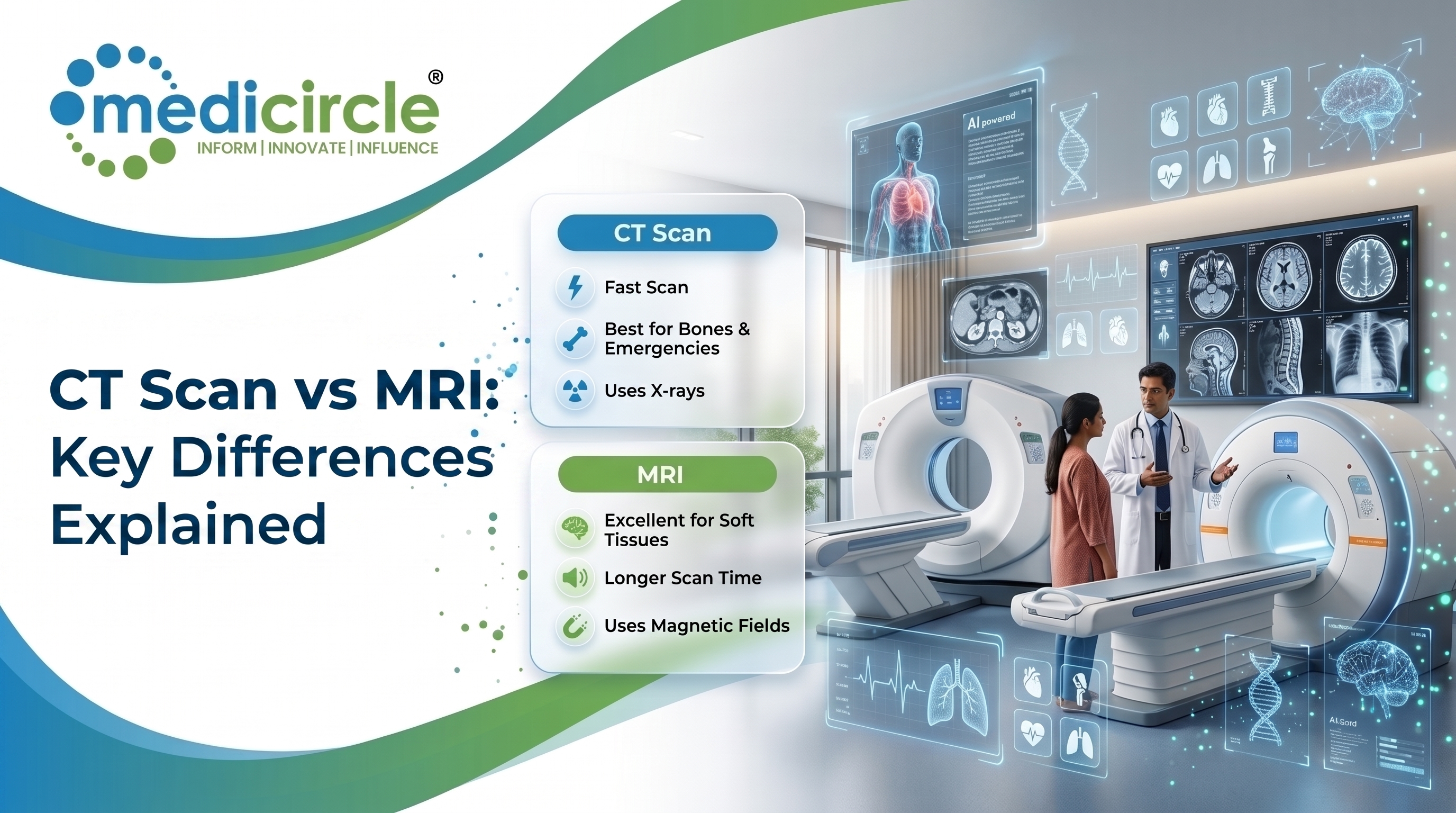
 CT scans use X-rays for fast, detailed imaging, ideal in emergencies, while MRIs use magnetic fields for superior soft tissue clarity. This article explains their differences, clinical uses, risks, and relevance to Indian patients.





Introduction
Medical imaging has transformed the way doctors diagnose and monitor disease. Among the most widely used diagnostic tools today are the CT scan and the MRI, two technologies that can reveal what is happening inside the human body without a single surgical incision. Yet for many patients in India, the question remains deeply confusing: what is the actual difference between a CT scan and an MRI, and which one should a doctor recommend?
This confusion is entirely understandable. Both tests involve lying inside a large machine. Both produce detailed internal images. And both are prescribed by doctors for conditions that range from headaches to cancer screening. But despite their surface similarities, the CT scan and the MRI are fundamentally different technologies, with different strengths, different risks, and very different clinical purposes.
Understanding these differences is not just useful for patients waiting in hospital corridors. In a country like India, where diagnostic imaging services are expanding rapidly across Tier 1 and Tier 2 cities, where out-of-pocket healthcare costs remain a significant concern, and where awareness of diagnostic procedures is still growing, informed patients make better decisions and receive better care.
Understanding the Two Technologies
What Is a CT Scan and How Does It Work?CT stands for computed tomography. A CT scan works by sending multiple X-ray beams through the body from different angles simultaneously. A rotating ring of detectors captures these beams and a computer processes them into detailed cross-sectional images, sometimes called slices, of the body's internal structures.
The entire scanning process is remarkably fast. Most CT scans are completed within five to ten minutes, and the actual imaging portion may take less than a minute. The machine itself looks like a large doughnut-shaped ring, and the patient lies on a table that moves smoothly through the center of this ring during the scan.
Because CT scans use X-rays, they produce ionizing radiation. The dose is higher than a standard chest X-ray but is kept to the minimum required for diagnostic quality. Modern CT machines in accredited hospitals and diagnostic centers across India are designed to minimize this dose without compromising image clarity.
What Is an MRI and How Does It Work?MRI stands for magnetic resonance imaging. Unlike a CT scan, an MRI does not use X-rays at all. Instead, it uses a powerful magnetic field combined with radio waves to create images of the body's internal structures.
When a patient enters the MRI scanner, the magnetic field causes hydrogen atoms in the body's water and fat molecules to align in a particular direction. Radio wave pulses are then sent through the body, causing these atoms to emit signals. A computer captures and translates these signals into highly detailed, three-dimensional images of tissues, organs, nerves, and blood vessels.
An MRI takes considerably longer than a CT scan. Depending on the body part being examined and whether contrast material is used, an MRI session typically lasts between twenty and sixty minutes. The machine produces loud thumping and knocking sounds during operation, and patients are usually offered earplugs or headphones to manage the noise.
Key Differences Between CT Scan and MRI
The following points highlight how these two technologies differ in meaningful and clinically relevant ways:
- Technology used: CT scans use ionizing radiation (X-rays), while MRI uses a magnetic field and radio waves.
- Speed: CT scans are significantly faster (five to ten minutes), while MRIs require twenty to sixty minutes or more.
- Image strength: MRI produces superior detail for soft tissues, nerves, muscles, and brain structures. CT scans excel at imaging bones, the chest, and abdominal organs quickly.
- Radiation: CT scans involve a small but measurable radiation dose. MRI involves no ionizing radiation whatsoever.
- Cost in India: CT scans are generally more affordable, ranging from approximately Rs. 1,500 to Rs. 8,000 depending on the body part and facility. MRI costs typically range from Rs. 4,000 to Rs. 20,000 or more for complex scans.
- Claustrophobia: MRI machines have a narrower enclosed bore, which can be challenging for patients with claustrophobia. CT scanners are more open in design.
- Metal implants: MRI cannot be performed on patients with certain metal implants, such as some pacemakers, cochlear implants, or older surgical clips. CT scans are generally safe for patients with implants.
When Doctors Recommend a CT Scan
CT scans are the imaging tool of choice in several common and critical clinical situations. Their speed makes them particularly valuable in emergencies, where rapid diagnosis can be the difference between life and serious harm.
In trauma settings, such as road accidents, which are unfortunately common across Indian roads, CT scans are used to quickly assess for internal bleeding, organ injury, skull fractures, or spinal damage. Emergency departments in larger hospitals in cities like Mumbai, Delhi, Bangalore, and Chennai rely heavily on CT scanners for exactly this reason.
CT scans are also widely used to detect lung conditions including infections, pulmonary embolism, and early-stage lung cancer. They provide excellent detail of the chest cavity and are a standard tool in oncology for staging cancer, monitoring treatment response, and detecting whether cancer has spread to the lungs, liver, or lymph nodes.
For abdominal conditions such as appendicitis, kidney stones, bowel obstructions, or abdominal aortic aneurysms, CT scans provide fast and reliable clarity. Coronary CT angiography has also emerged as a powerful non-invasive tool for evaluating heart artery blockages, increasingly available in cardiac centers across India.
When Doctors Recommend an MRI
MRI is the preferred imaging modality when soft tissue detail is the priority and when radiation-free imaging is a clinical advantage.
Neurological conditions represent one of the most important use cases for MRI in India. Brain tumors, strokes, multiple sclerosis, epilepsy, and infections such as meningitis or encephalitis are all assessed with MRI because of its superior ability to differentiate between healthy brain tissue and abnormal tissue. The ICMR has noted the rising burden of neurological disorders in India, making brain MRI an increasingly utilized diagnostic tool.
Spinal conditions such as herniated discs, spinal cord compression, and back pain of uncertain origin are commonly evaluated through spinal MRI. Orthopedic injuries including torn ligaments, rotator cuff tears, meniscal injuries, and joint inflammation are also far better visualized on MRI than on CT or X-ray.
In women's health, MRI is used for breast cancer evaluation, particularly in high-risk cases where mammography alone may not be sufficient. Prostate cancer evaluation, liver lesion characterization, and uterine and ovarian assessment also benefit from MRI's detail.
For pregnant women who require imaging, MRI is the safer alternative to CT because it does not expose the developing fetus to ionizing radiation. Doctors typically exercise caution and recommend MRI only when clinically essential, generally avoiding it during the first trimester.
Risks and Safety Considerations
Both CT scans and MRIs are widely regarded as safe diagnostic procedures when ordered appropriately and performed in accredited facilities. However, understanding their respective risk profiles allows patients to have more informed conversations with their doctors.
The primary risk associated with CT scans is ionizing radiation. A single CT scan delivers a radiation dose that, for most adults, represents an acceptably small risk. However, cumulative radiation exposure from multiple scans over time is a genuine consideration, particularly for children, young adults, and patients who require frequent imaging. In India, the Atomic Energy Regulatory Board (AERB) oversees radiation safety standards for diagnostic facilities, and accredited centers are required to follow dose optimization protocols.
Contrast dye is sometimes used in both CT and MRI scans to improve image quality. CT scans use iodine-based contrast, which can cause allergic reactions in a small number of patients and may affect kidney function in those with pre-existing renal disease. MRI contrast uses gadolinium-based agents, which carry a different risk profile and are also used with caution in patients with kidney disease.
MRI carries no radiation risk, which is one of its most significant advantages for pediatric patients and for women of childbearing age. However, patients with certain metallic implants must inform their doctors before an MRI is scheduled. Ferromagnetic materials in the body can move or heat in response to the MRI's magnetic field. Modern MRI facilities screen patients carefully through detailed pre-scan questionnaires to identify any contraindications.
The enclosed design of MRI scanners can trigger claustrophobia or anxiety in some patients. Sedation is available for patients who need it, and open MRI machines, though offering lower magnetic field strength, are available at select centers for patients who find standard MRI machines uncomfortable.
Contrast Agents: An Important Distinction
Both modalities offer the option of using contrast agents to enhance visibility of specific structures or abnormalities. However, the contrast agents used in CT and MRI are chemically different and serve different imaging purposes.
CT contrast uses iodine-based solutions, which are typically administered intravenously before or during the scan. Patients with known iodine allergies should inform their radiologist in advance, as alternative protocols or pre-medications can be arranged.
MRI contrast uses gadolinium-based contrast agents (GBCAs), which enhance visibility of blood vessels, tumors, inflammation, and areas of abnormal blood-brain barrier breakdown. These agents carry a risk called nephrogenic systemic fibrosis (NSF) in patients with severely reduced kidney function, and their use in such cases requires careful clinical judgment.
CT Scan vs MRI in the Indian Healthcare Context
The expansion of diagnostic imaging infrastructure in India over the past decade has been substantial. According to NITI Aayog reports and various healthcare sector analyses, the availability of CT scanners and MRI machines has increased significantly in both urban hospitals and district-level health centers. Government initiatives such as the Pradhan Mantri Jan Arogya Yojana (PM-JAY) under Ayushman Bharat have extended coverage for diagnostic procedures including CT and MRI to millions of low-income households.
However, access disparities persist. In many rural and semi-urban areas, CT scanners may be available, but MRI machines remain limited to larger district hospitals or private diagnostic chains. Patients traveling from smaller towns to cities for MRI scans is still a common reality. The growth of portable and mobile MRI units, as well as government investment in AIIMS regional campuses and upgraded district hospitals, represents a meaningful step toward bridging this gap.
Cost remains a significant factor in patient decision-making. While insurance coverage under PM-JAY and corporate health plans has improved affordability for many, a large proportion of India's population continues to pay out-of-pocket for diagnostic imaging. Understanding what each modality costs and what clinical value it provides helps patients make decisions that balance clinical need with financial reality.
Frequently Asked Questions
Q1: What is the main difference between a CT scan and an MRI?
A CT scan uses X-rays to create cross-sectional images of the body, making it ideal for bones, organs, and emergency assessments. An MRI uses magnetic fields and radio waves to produce highly detailed images of soft tissues, nerves, and organs without radiation exposure. The choice between the two depends on what part of the body needs to be examined and for what clinical reason.
Q2: Which is safer, CT scan or MRI?
MRI is generally considered safer for repeated use because it does not involve ionizing radiation. CT scans carry a small radiation dose but are still considered safe for most patients when clinically necessary. Both modalities are regarded as safe when performed in accredited facilities under appropriate clinical guidance.
Q3: Why is MRI more expensive than CT scan in India?
MRI machines are significantly more costly to install, operate, and maintain compared to CT scanners. The scanning procedure itself takes considerably longer, which limits the number of patients a facility can scan per day. These factors together contribute to the higher cost of MRI across Indian hospitals and diagnostic centers.
Q4: Can a pregnant woman undergo a CT scan or MRI?
Pregnant women are generally advised to avoid CT scans due to ionizing radiation exposure to the developing fetus. MRI is considered a safer alternative during pregnancy and is used when clinical necessity demands it. Doctors typically recommend avoiding MRI during the first trimester unless the benefit clearly outweighs any potential risk.
Q5: How long does a CT scan take compared to an MRI?
A CT scan is much faster, typically completing in five to ten minutes, with the actual imaging taking under one minute. An MRI typically takes between twenty and sixty minutes depending on the body part and whether contrast is used. This speed difference is one reason CT scans are preferred in emergency settings.
Resources
- Indian Council of Medical Research (ICMR): Guidelines and publications on diagnostic radiology standards and disease burden in India
- World Health Organization (WHO): India Country Office reports on non-communicable disease diagnostics and imaging access
- Atomic Energy Regulatory Board (AERB), Government of India: Radiation safety regulations for diagnostic imaging facilities
- Ministry of Health and Family Welfare (MoHFW): Information on Ayushman Bharat PM-JAY coverage and diagnostic benefits
- NITI Aayog: Reports on healthcare infrastructure expansion including diagnostic imaging in India
Interlinking Keywords:
MRI scan procedure India, CT scan cost India, contrast dye CT scan, brain MRI diagnosis, radiation safety diagnostic imaging, Ayushman Bharat diagnostic coverage, medical imaging types, radiology in India
Last medically reviewed by:
Medicircle Editorial Health Desk on July 8, 2026
Medical Disclaimer:
This article is intended for general informational and educational purposes only. It does not constitute medical advice, diagnosis, or treatment. Always consult a qualified and licensed medical professional for any concerns related to your health or before making decisions about diagnostic tests. Medicircle does not recommend specific diagnostic centers, treatments, or procedures.






.jpeg)
.jpg)
.jpg)
.jpg)
.jpg)
.jpg)
.jpg)






.jpeg)

.jpeg)


.png)