 Monday, November 02, 2026
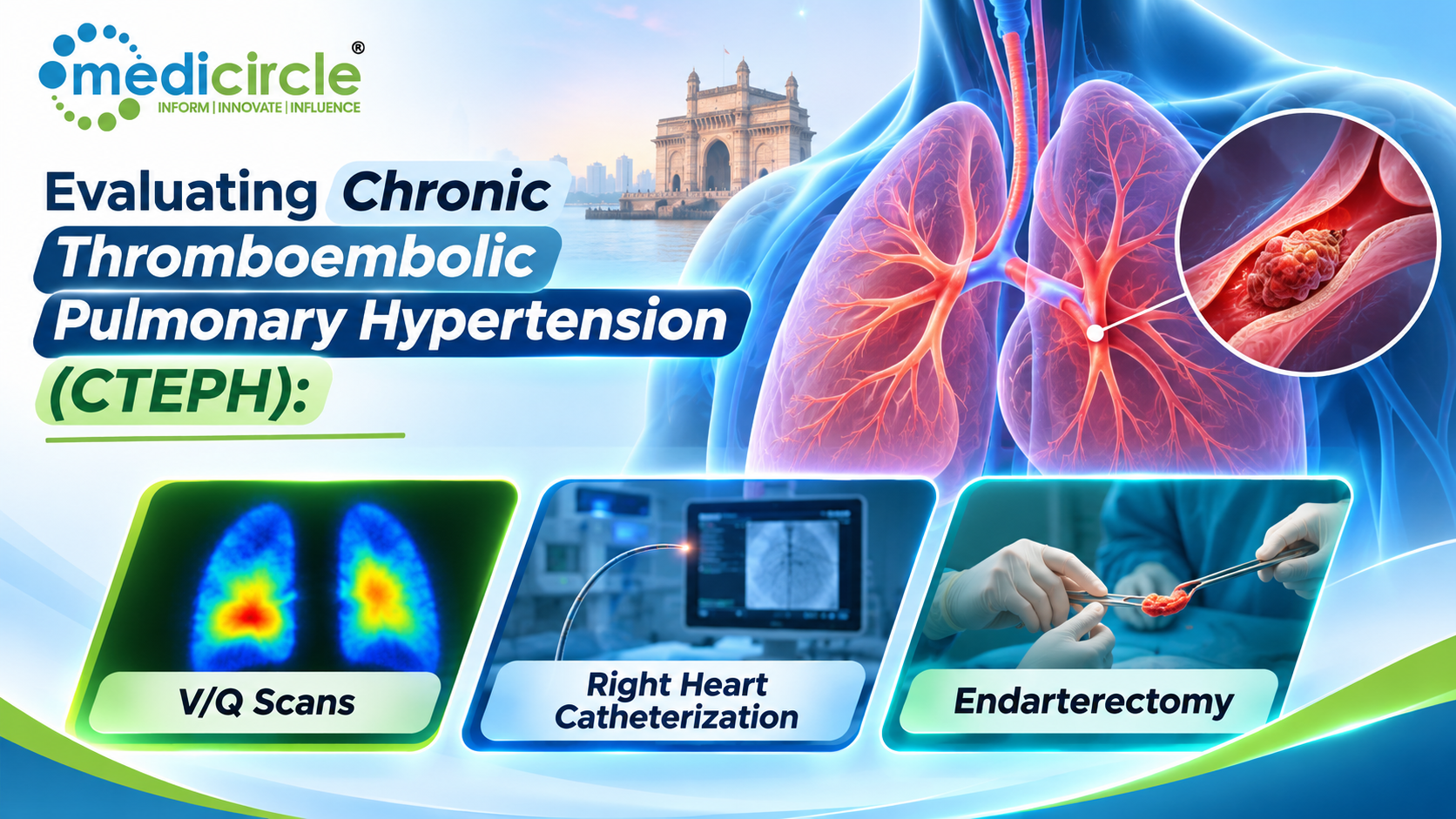
 Evaluating Chronic Thromboembolic Pulmonary Hypertension (CTEPH) involves V/Q scans to detect pulmonary blood flow abnormalities and right heart catheterization to confirm pulmonary hypertension and assess its severity. Pulmonary endarterectomy offers a potentially curative treatment for eligible patients by removing chronic blood clots, improving circulation, and restoring heart and lung function.





Chronic Thromboembolic Pulmonary Hypertension (CTEPH) is a distinct form of pulmonary hypertension categorized as Group 4 Pulmonary Hypertension under World Health Organization (WHO) classifications. It is characterized by the incomplete resolution and subsequent fibrothrombotic transformation of single or recurrent pulmonary emboli. Over time, these organized, endothelialized fibrotic obstructions occlude major, lobar, segmental, and subsegmental pulmonary arteries, triggering secondary small-vessel remodeling, progressive pulmonary vascular resistance (PVR), and eventual right heart failure.
Unlike pulmonary arterial hypertension (PAH / Group 1), CTEPH represents a potentially curable etiology of pulmonary hypertension, provided it is recognized promptly and evaluated systematically through ventilation-perfusion scintigraphy, invasive hemodynamics, and expert surgical assessment.
The Diagnostic Anchor: Ventilation-Perfusion (V/Q) Scintigraphy
A major contributor to delayed CTEPH diagnoses is reliance on standard non-contrast CT or conventional CT Pulmonary Angiography (CTPA) alone. While CTPA is essential for anatomic mapping later in the workup, ventilation-perfusion (V/Q) lung scintigraphy remains the frontline screening modality of choice due to its unmatched sensitivity.
Sensitivity and Screening Value- Diagnostic Sensitivity: A normal or very low-probability planar V/Q scan rules out CTEPH with a sensitivity exceeding 96% to 97% and a negative predictive value approaching 100%. CTPA, while highly specific for central proximal disease, frequently underdetects eccentric webs, bands, and subsegmental micro-vascular lesions when interpreted outside expert CTEPH centers.
- Characteristic Pattern: The hallmark of CTEPH is the presence of mismatched segmental or subsegmental perfusion defects—anatomical segments with preserved ventilation but absent or sharply reduced perfusion in regions matching vascular anatomy.
Where available, 3D single-photon emission computed tomography combined with non-contrast CT (V/Q SPECT/CT) has largely superseded planar scintigraphy. SPECT/CT eliminates anatomical overlap, improves attenuation correction, detects subsegmental lesions with greater precision, and directly co-registers functional perfusion voids with underlying parenchymal morphology.
Hemodynamic Confirmation: Right Heart Catheterization (RHC)
Every patient with suspected CTEPH and a positive or indeterminate V/Q scan must undergo a standardized resting Right Heart Catheterization (RHC) to confirm precapillary pulmonary hypertension and quantify the hemodynamic burden.
Diagnostic CriteriaUnder current international consensus guidelines (including ESC/ERS criteria), CTEPH is hemodynamically defined at rest in a patient with chronic non-clearing thromboembolic vascular obstructions after at least 3 months of therapeutic anticoagulation by:
- Mean Pulmonary Arterial Pressure (mPAP): > 20 mmHg
- Pulmonary Arterial Wedge Pressure (PAWP): ≤ 15 mmHg (confirming a precapillary rather than postcapillary etiology)
- Pulmonary Vascular Resistance (PVR): > 2 Wood Units (WU) (or > 160 dyn·s·cm⁻⁵)
(Note: Patients exhibiting typical chronic thromboembolic vascular lesions and exercise limitation without resting pulmonary hypertension—mPAP ≤ 20 mmHg with PAWP ≤ 15 mmHg—are classified as having Chronic Thromboembolic Pulmonary Disease (CTEPD) without resting PH).
Clinical Utility of Hemodynamic Profiling- Right Ventricular Function Assessment: Measuring right atrial pressure (RAP), mixed venous oxygen saturation (SvO_2), and cardiac output (CO) via thermodilution or the direct Fick method provides direct insight into right ventricular compensation or failure.
- Risk Stratification: Extremely elevated PVR (e.g., > 1,000–1,200 dyn·s·cm⁻⁵) and high RAP signal severe right ventricular afterload and disease chronicity, guiding perioperative management and inotropic support strategies.
Defining Operability: Anatomical Mapping and Patient Selection
The identification of CTEPH on V/Q scanning and RHC triggers an immediate multidisciplinary evaluation to determine whether the patient is a candidate for Pulmonary Endarterectomy (PEA). This assessment involves dual-source or high-resolution CTPA and invasive biplane Digital Subtraction Pulmonary Angiography (DSA).
Angiographic signs indicative of chronic thromboembolic disease include:
- Vascular webs, bands, and slits
- Pouch defects and complete vascular cutoffs
- Irregular vessel wall contours and post-stenotic dilatations
- Sparing and hyper-perfusion of unobstructed vascular beds
Operability is determined not strictly by hemodynamic cutoffs, but by the spatial concordance between the patient’s hemodynamic elevation and surgically accessible vascular blockages, weighed against comorbidities and performance status.
The Definitive Cure: Pulmonary Endarterectomy (PEA)
Pulmonary Endarterectomy (PEA)—sometimes called Pulmonary Thromboendarterectomy (PTE)—is the standard of care and primary curative therapy for operable CTEPH.
Key Technical Aspects of PEA- True Endarterectomy vs. Embolectomy: PEA is not a simple extraction of fresh thrombus. It requires a meticulous surgical dissection within the media layer of the pulmonary arterial wall to strip away chronic, organized, fibrous scar tissue down to the segmental and subsegmental branching vessels.
- Median Sternotomy and Cardiopulmonary Bypass (CPB): Both pulmonary arteries are accessed through a midline approach.
- Deep Hypothermic Circulatory Arrest (DHCA): To visualize and clear delicate subsegmental vessels without bleeding from extensive bronchial arterial collaterals, the patient’s core temperature is lowered to deep hypothermia (typically 18°C–20°C). The surgeon operates during controlled, intermittent periods of circulatory arrest (usually 20-minute intervals).
In high-volume specialized surgical centers, in-hospital mortality for PEA is below 2% to 3%. A successful endarterectomy results in:
- Immediate, dramatic reduction in pulmonary vascular resistance and mPAP
- Decompression and reverse remodeling of the enlarged right ventricle
- Normalization of cardiac index and functional capacity (often moving patients from WHO Functional Class III/IV back to Class I/II)
- Discontinuation of supplemental oxygen and long-term improvement in quality of life
For patients deemed inoperable due to extreme distal/microvascular lesions, high frailty, or significant residual pulmonary hypertension post-PEA:
- Balloon Pulmonary Angioplasty (BPA): A catheter-based interventional approach using sequential balloon dilations to fracture intraluminal webs and bands in distal segmental/subsegmental vessels.
- Targeted Medical Therapy: Soluble guanylate cyclase (sGC) stimulators (such as Riociguat) are approved to reduce PVR and improve exercise capacity in inoperable CTEPH or persistent/recurrent post-operative PH.
Frequently Asked Questions (FAQs)
1. What is Chronic Thromboembolic Pulmonary Hypertension (CTEPH)?CTEPH is a progressive form of pulmonary hypertension (WHO Group 4) caused by the chronic persistence and fibrotic remodeling of pulmonary emboli. These organized fibrous tissues obstruct the pulmonary arterial tree, raising pulmonary vascular resistance and eventually leading to right-sided heart failure.
2. Why is a V/Q scan preferred over CTPA for initial CTEPH screening?A ventilation-perfusion (V/Q) scan is significantly more sensitive (over 96%) than CT pulmonary angiography for identifying chronic subsegmental thromboembolic lesions. While CTPA is effective for visualizing central anatomy, standard multi-detector CT can miss subtle peripheral webs and bands, making V/Q the mandatory screening test.
3. What are the hemodynamic criteria for diagnosing CTEPH on a right heart catheterization?Following at least 3 months of therapeutic anticoagulation, CTEPH is confirmed when RHC demonstrates a resting mean pulmonary arterial pressure (mPAP) > 20 mmHg, a normal pulmonary arterial wedge pressure (PAWP) ≤ 15 mmHg, and a pulmonary vascular resistance (PVR) > 2 Wood Units (> 160 dyn·s·cm⁻⁵).
4. What is the difference between an acute pulmonary embolism and CTEPH?An acute pulmonary embolism consists of a fresh, soft, red blood clot that can be lysed medically or surgically retrieved via standard embolectomy. CTEPH represents chronic, non-resolving organized scar tissue that has incorporated into the vessel wall intima and media, requiring a surgical endarterectomy plane for removal.
5. What is Pulmonary Endarterectomy (PEA)?Pulmonary Endarterectomy is a complex open-heart surgery designed to treat CTEPH. Performed via median sternotomy under cardiopulmonary bypass and deep hypothermic circulatory arrest, the surgeon dissects into the vessel wall to peel out chronic fibrotic obstructions from the main, lobar, segmental, and subsegmental pulmonary arteries.
6. Why is Deep Hypothermic Circulatory Arrest (DHCA) required during PEA?In CTEPH, systemic-to-pulmonary bronchial arterial collaterals proliferate substantially. Even on full cardiopulmonary bypass, retrograde bronchial collateral flow floods the surgical field with blood. DHCA provides a completely bloodless, motionless field, enabling the surgeon to visualize and cleanly dissect delicate subsegmental vessel branches without tearing the arterial wall.
7. Can CTEPH occur without a documented history of acute deep vein thrombosis (DVT) or pulmonary embolism (PE)?Yes. Up to 25% to 30% of patients diagnosed with CTEPH have no prior clinical documentation of an acute deep vein thrombosis or pulmonary embolism. Chronic clot formation can proceed subclinically or be misattributed to asthma, COPD, or deconditioning until progressive dyspnea manifests.
8. What are the common symptoms of CTEPH?The most frequent presenting symptom is progressive exertional dyspnea. Other symptoms include chronic fatigue, exercise intolerance, lightheadedness, syncope upon exertion, chest tightness, peripheral lower-extremity edema, and signs of right heart strain.
9. What treatment options exist if a patient is deemed inoperable for PEA?Patients with distal microvascular disease or severe co-morbidities who are not candidates for PEA can be managed with targeted medical therapy (primarily the soluble guanylate cyclase stimulator Riociguat) and sequential Balloon Pulmonary Angioplasty (BPA), an interventional catheter-based approach that mechanically disrupts distal webs and rings.
10. Does a successful pulmonary endarterectomy cure the condition permanently?In a large proportion of patients with operable, proximal-to-segmental disease, PEA provides a functional cure, returning resting pulmonary pressures and right ventricular function to normal or near-normal levels. However, patients require lifelong therapeutic anticoagulation to prevent recurring thromboembolic events and routine clinical follow-up to monitor for residual or recurrent pulmonary hypertension.





-23-300x250.png)