Sunday, October 25, 2026
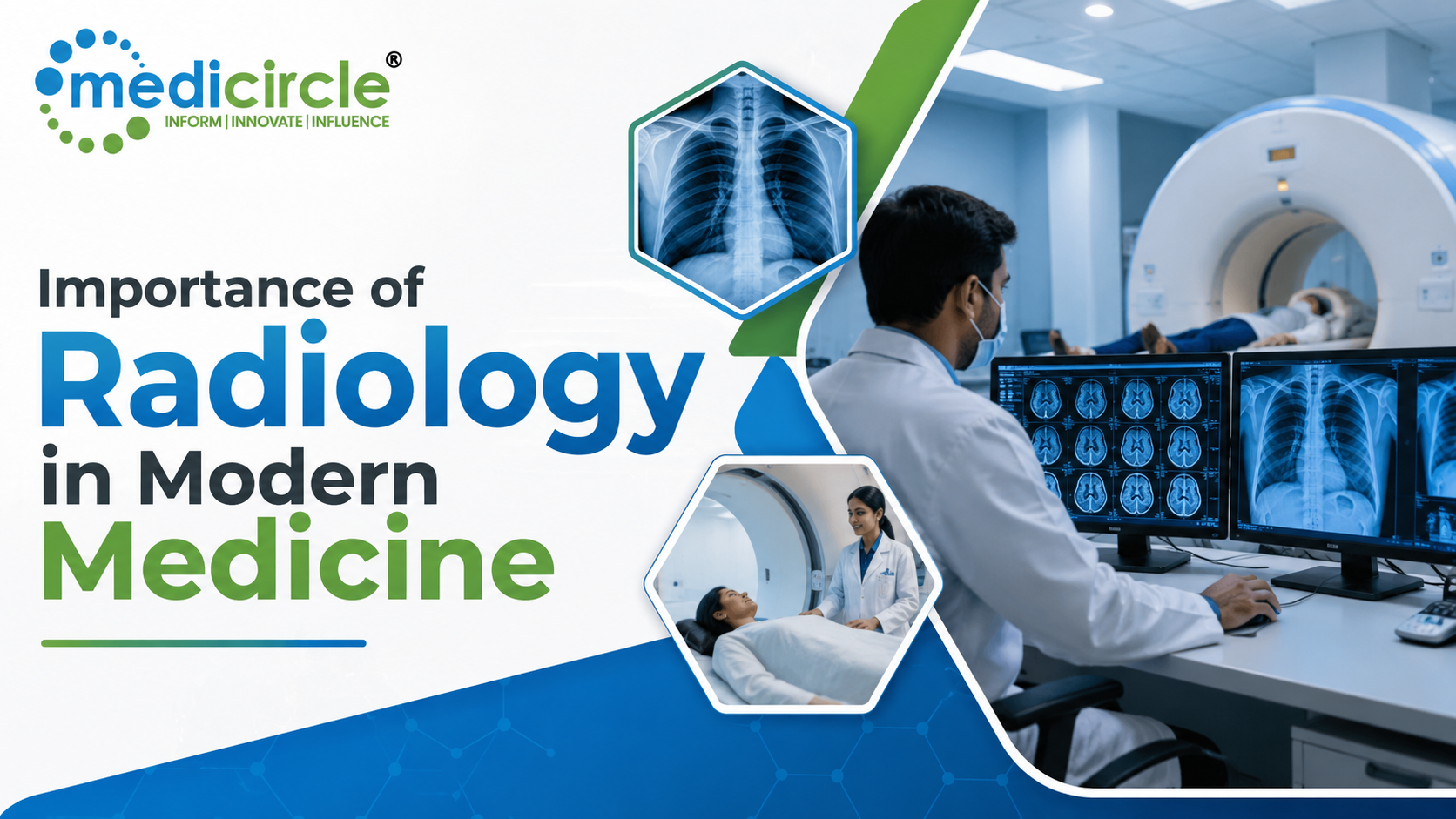
 Radiology is a foundational medical specialty that enables early disease detection, precise treatment planning, and minimally invasive procedures, making it indispensable to modern Indian healthcare delivery.





Introduction
Modern medicine depends on the ability to see what cannot be seen with the naked eye. Whether a doctor is evaluating a patient with chest pain, a child with a suspected bone fracture, a woman with a breast lump, or a person with neurological symptoms, the next step almost always involves medical imaging. Radiology is the medical specialty that makes this possible.
Radiology refers to the use of radiant energy and imaging technologies to diagnose and, increasingly, to treat a wide range of medical conditions. It has transformed the practice of medicine in every clinical discipline, from oncology and cardiology to orthopaedics, neurology, and emergency care. Without radiology, a significant portion of modern diagnosis would simply not be possible.
In India, where the healthcare system is managing a growing burden of non-communicable diseases alongside existing infectious disease challenges, the role of radiology has never been more important. As the country continues to invest in healthcare infrastructure under initiatives such as Ayushman Bharat and the Ayushman Bharat Digital Mission (ABDM), radiology services are becoming more accessible across both urban centres and Tier 2 and Tier 3 cities. Understanding the importance of radiology is therefore relevant not just for medical professionals but for every patient, hospital administrator, policymaker, and healthcare stakeholder in India.
Understanding the Basics of Radiology
Radiology as a formal medical specialty traces its origin to 1895, when Wilhelm Conrad Roentgen discovered X-rays and produced the first radiographic images. Since then, the field has expanded dramatically to encompass a wide range of imaging technologies that form the diagnostic foundation of contemporary clinical practice.
At its core, radiology is divided into two primary branches.
Diagnostic Radiology focuses on producing and interpreting images of the human body to identify disease, injury, or abnormality. This branch includes the most commonly used imaging tools such as X-ray, CT scan, MRI, ultrasound, mammography, and nuclear medicine imaging including PET scans.
Interventional Radiology uses real-time imaging to guide minimally invasive procedures inside the body. Rather than relying solely on imaging for diagnosis, interventional radiologists use imaging tools as a roadmap to perform treatments such as biopsies, drainage procedures, angioplasties, tumour ablations, and embolisations.
The specialists who practise radiology, known as radiologists, are fully qualified medical doctors who undergo several additional years of specialized training in interpreting medical images and correlating imaging findings with clinical presentations. They work in close collaboration with every other clinical specialty and serve as a critical link in the chain of patient care.
The Core Imaging Modalities and What They Reveal
The power of radiology lies in its diverse toolkit. Each imaging modality has distinct physical principles, clinical applications, and strengths that make it valuable in specific situations.
X-ray remains the most widely used and accessible imaging tool. It is particularly effective for evaluating bone fractures, joint conditions, dental problems, and lung pathology, including pneumonia, tuberculosis, and pleural effusions. In India, where tuberculosis continues to be a major public health concern, chest X-rays remain a primary screening and diagnostic tool.
Computed Tomography (CT) Scans use multiple X-ray projections processed by a computer to generate detailed cross-sectional images of the body. CT scans are invaluable in emergency settings for detecting internal bleeding, organ injuries, pulmonary embolism, and stroke. They are also extensively used in cancer staging, surgical planning, and monitoring treatment response.
Magnetic Resonance Imaging (MRI) uses magnetic fields and radiofrequency waves rather than ionizing radiation. MRI offers superior soft tissue contrast and is the preferred investigation for brain and spinal cord conditions, musculoskeletal injuries, liver disease, and gynaecological conditions. It has revolutionized the fields of neurology and orthopaedics.
Ultrasound uses high-frequency sound waves to create real-time images of organs and soft tissues. It is safe, portable, and widely used in obstetrics for foetal monitoring, as well as for evaluating abdominal organs, thyroid, and guiding procedures such as biopsies. In India, point-of-care ultrasound is expanding access to diagnostic imaging even in primary health settings.
PET Scans and Nuclear Medicine use small amounts of radioactive tracers to visualize metabolic activity inside the body. These are particularly critical in oncology for detecting cancer spread, evaluating treatment response, and identifying recurrence.
Mammography is a specialized low-dose X-ray designed specifically for breast tissue evaluation. Given the rising incidence of breast cancer in India, with studies indicating that it is now the most common cancer among women in urban areas, mammography-based screening programmes have become a public health priority.
The Clinical Importance of Radiology Across Medical Specialties
One of the most compelling aspects of radiology is its indispensability across every clinical specialty. There is virtually no area of medicine that functions independently of imaging.
In oncology, radiology is involved at every stage of cancer care. Imaging detects tumours, defines their location and extent, assists in biopsy planning, guides radiation therapy, and monitors treatment response. Interventional radiology has added minimally invasive treatment options that reduce the need for major surgery.
In cardiology, CT coronary angiography, echocardiography, and cardiac MRI provide non-invasive assessments of heart structure, function, and blood vessel health. These tools are central to diagnosing and managing coronary artery disease, which is now among the leading causes of death in India.
In neurology, MRI and CT scans are the primary tools for diagnosing stroke, brain tumours, multiple sclerosis, epilepsy, and degenerative brain conditions. The speed and accuracy of CT in identifying haemorrhagic stroke have directly contributed to better outcomes in emergency neurology.
In orthopaedics, X-rays and MRI guide fracture management, joint replacement planning, and the evaluation of ligament and tendon injuries. In obstetrics, ultrasound is the cornerstone of antenatal monitoring. In gastroenterology, CT and MRI enterography evaluate inflammatory bowel disease, liver conditions, and pancreatic pathology.
Emergency medicine perhaps demonstrates the most acute dependence on radiology. In the critical first hour of a medical emergency, whether trauma, stroke, or acute abdomen, imaging findings directly determine the clinical pathway and can mean the difference between life and disability or death.
Radiology's Role in Treatment Planning and Monitoring
The importance of radiology extends far beyond initial diagnosis. It plays an equally vital role in planning and monitoring treatment across multiple disciplines.
Surgeons rely on detailed CT and MRI images to plan operations precisely, mapping the location, size, and relationship of a tumour or anatomical abnormality to surrounding structures before making a single incision. Radiation oncologists use PET-CT imaging to define target volumes for radiotherapy with millimetre-level accuracy, sparing healthy tissue while maximizing the dose delivered to cancer.
After treatment, whether surgery, chemotherapy, or radiation, radiology tracks the patient's response. Serial imaging helps oncologists determine whether a treatment is working, allows early detection of recurrence, and enables timely modification of the treatment plan.
Interventional radiology has further extended this role into the realm of active treatment. Procedures such as radiofrequency ablation and microwave ablation destroy tumours from within, guided by real-time imaging. Transarterial chemoembolisation delivers chemotherapy directly to a liver tumour through its blood supply while simultaneously blocking that supply. These procedures offer effective alternatives for patients who are not surgical candidates.
The State of Radiology in India: Challenges and Opportunities
India presents a unique and complex landscape for radiology. On one hand, major metropolitan hospitals in cities such as Mumbai, Delhi, Bengaluru, Chennai, and Hyderabad are equipped with advanced imaging infrastructure and internationally trained radiologists. On the other hand, rural India and smaller towns continue to face significant gaps in access to basic radiology services.
According to available data, India has a severe shortage of radiologists relative to its population, with estimates suggesting fewer than 15,000 qualified radiologists to serve a population of over 1.4 billion people. This disparity is most pronounced in public health facilities and rural districts.
Teleradiology has emerged as a significant solution to this challenge. By transmitting digital images over secure networks to remote radiologists for interpretation, teleradiology has made specialist-level diagnostic reporting available to district hospitals and primary care centres that lack an on-site radiologist. Several Indian startups and established healthcare companies are now operating teleradiology platforms that serve hundreds of hospitals across the country.
The government's push toward digital health infrastructure under ABDM is expected to further integrate radiology into the broader healthcare ecosystem, enabling seamless sharing of imaging reports as part of a patient's unified health record. Additionally, National Health Policy guidelines have emphasized strengthening diagnostic services, including radiology at district hospital levels under the National Health Mission.
Artificial Intelligence and the Future of Radiology
Artificial intelligence is rapidly emerging as one of the most transformative forces in radiology worldwide, and India is not far behind in exploring its potential.
AI algorithms trained on large datasets of medical images can now detect abnormalities in chest X-rays, identify diabetic retinopathy from fundus photographs, flag suspicious lesions in mammograms, and assist in measuring and characterizing tumours on CT scans with a speed and consistency that complements radiologist expertise. In high-volume settings such as Indian public hospitals, where a single radiologist may need to review hundreds of images per day, AI-assisted tools hold enormous promise for improving efficiency and reducing errors.
Several Indian healthcare technology companies are developing AI-powered radiology tools calibrated to Indian disease patterns and imaging equipment commonly found in Indian hospitals. This is particularly important because AI models trained exclusively on Western patient populations may not perform optimally for Indian patients, who may present with different disease patterns, body compositions, and imaging characteristics.
The integration of AI with teleradiology platforms creates a compelling model for expanding access to quality diagnostic imaging across India at scale, aligning with the broader goals of equitable healthcare delivery.
The Radiologist as a Clinical Partner
A common misconception is that radiology is purely a technical service where images are produced and reports are filed. In reality, the modern radiologist is a clinical partner whose expertise shapes diagnosis, guides treatment, and contributes meaningfully to multidisciplinary patient care teams.
Radiologists participate in tumour boards alongside oncologists and surgeons, contribute to trauma response protocols, collaborate with interventional cardiologists during complex cardiac procedures, and consult on difficult diagnostic cases across all specialties. Their ability to correlate imaging findings with clinical history, laboratory results, and patient symptoms is what distinguishes expert radiological interpretation from automated image analysis.
For patients, direct interaction with a radiologist, whether before an MRI to explain the procedure or after an interventional procedure to discuss the findings, can significantly improve the care experience. As Indian hospitals increasingly adopt patient-centred care models, the visible and communicative role of the radiologist is becoming more valued.
Platforms such as Medicircle play an important role in amplifying the voice and expertise of radiologists and other medical specialists, helping them share knowledge with a broader public audience and contributing to greater health literacy around radiology and diagnostic imaging in India.
Frequently Asked Questions
Q1: What is the importance of radiology in healthcare?
Radiology enables non-invasive diagnosis of diseases through imaging techniques such as X-ray, MRI, CT scan, and ultrasound. It allows early detection, accurate disease staging, treatment planning, and monitoring of patient progress, making it one of the most critical pillars of modern medicine.
Q2: What are the different types of radiology?
Radiology is broadly divided into diagnostic radiology and interventional radiology. Diagnostic radiology includes X-rays, CT scans, MRI, ultrasound, PET scans, and mammography. Interventional radiology uses imaging to guide minimally invasive procedures such as biopsies, angioplasties, and tumour ablations.
Q3: How is radiology used in cancer care in India?
In India, radiology plays a vital role in cancer detection and management. PET scans and CT scans help in staging cancers, while mammography supports early breast cancer detection. Interventional radiology techniques such as tumour ablation and chemoembolisation offer minimally invasive treatment alternatives for cancer patients.
Q4: Is radiology safe for patients?
Most radiology investigations are safe. Ultrasound and MRI do not use ionizing radiation and are considered completely safe. X-rays and CT scans involve low doses of radiation and are justified when the clinical benefit clearly outweighs the risk. Radiologists are trained in radiation protection to ensure patient safety at all times.
Q5: What is the difference between a radiologist and a radiology technician?
A radiologist is a medically qualified doctor who specializes in interpreting medical images and providing clinical diagnoses. A radiology technician or radiographer is a trained healthcare professional who operates imaging equipment, positions patients, and produces the images that the radiologist subsequently interprets.
Resources
- Indian Council of Medical Research (ICMR): Guidelines, publications, and research data on cancer incidence, diagnostic imaging standards, and disease burden in India.
- World Health Organization (WHO), India Country Office: Reports and frameworks on strengthening diagnostic and imaging services in low- and middle-income country health systems.
- Ministry of Health and Family Welfare, Government of India (mohfw.gov.in): National Health Policy documents, National Health Mission frameworks, and Ayushman Bharat programme guidelines relevant to diagnostic radiology.
- Ayushman Bharat Digital Mission (abdm.gov.in): Information on the national digital health infrastructure including standards for imaging data integration into unified health records.
- PubMed, National Library of Medicine (pubmed.ncbi.nlm.nih.gov): Peer-reviewed research articles on radiology's evolving role in clinical medicine, AI in radiology, teleradiology, and Indian healthcare studies.
Interlinking Keywords:
diagnostic imaging in India, interventional radiology, radiology types, teleradiology in India, role of radiologist, AI in radiology, Ayushman Bharat healthcare, cancer diagnosis imaging, MRI scan India, CT scan uses
Last medically reviewed by:
Editorial and Medical Advisory Team, Medicircle on August 1, 2025
Medical Disclaimer:
This article is intended for general awareness and educational purposes only. It does not constitute medical advice, diagnosis, or a treatment recommendation. Readers should always consult a qualified and registered medical professional for any health-related concerns, imaging requirements, or clinical decisions. Medicircle does not promote any specific diagnostic facility, imaging brand, or medical service provider.





-23-300x250.png)

.jpeg)
.jpg)
.jpg)
.jpg)
.jpg)
.jpg)
.jpg)
.jpg)
.jpg)
.jpg)