 Thursday, October 08, 2026
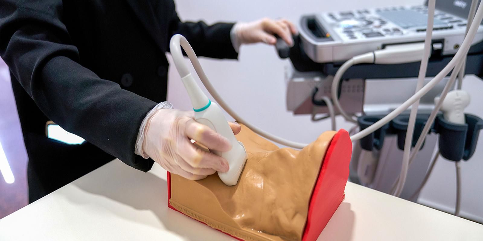
 Moscow has expanded its innovative medical training capabilities with the development of two new realistic phantoms: a stomach and a pulsating arterial vessel, both possessing unique characteristics.




Moscow has expanded its innovative medical training capabilities with the development of two new realistic phantoms: a stomach and a pulsating arterial vessel, both possessing unique characteristics. These phantoms are used by doctors to hone their skills in complex procedures and ultrasound diagnostics, as well as to monitor the quality of CT and MRI scans. With these additions, Moscow scientists have now created a total of 14 highly realistic models, which are employed for advanced doctor training and precise diagnostic equipment calibration. This was confirmed by Anastasia Rakova, Deputy Mayor of Moscow for Social Development.
Moscow is consistently implementing innovative technologies not only to advance diagnostic and treatment capabilities, but also to modernize its physician training systems. A key example of this initiative is the ongoing development of highly realistic anatomical phantoms—precise models that replicate the unique properties and characteristics of human tissue and physiology. These phantoms provide clinicians with essential tools to practice ultrasound diagnostics, verify the quality of CT and MRI scans, and refine their techniques for complex medical procedures. To date, scientists in Moscow have created a suite of 14 distinct models. As Anastasia Rakova explained, "The newest of these are the stomach phantom and the pulsating arterial vessel phantom."
The stomach phantom is modeled as a hollow organ featuring a five-layer wall, with each stratum possessing distinct ultrasonic reflectance properties. This construction generates imaging results nearly identical to those obtained from a real human stomach. The innermost layer, which mimics the gastric mucosa, incorporates a realistically folded anatomical structure. Additionally, models of neoplasms are embedded at varying depths within these layers. This design makes the phantom an invaluable training tool for practicing endoscopy (gastroscopy), mastering ultrasound diagnostics, and refining biopsy techniques.
During an ultrasound-guided gastric biopsy simulation, the clinician must precisely navigate the needle into the targeted layer to reach the simulated tumor. Given that each wall layer is only approximately one to three millimeters thick, the procedure demands extreme accuracy to avoid puncturing an incorrect layer or penetrating entirely through the organ wall.
The pulsating arterial vessel phantom is a dynamic model designed to mimic the precise pulse wave within a specific section of a blood vessel. It comprises two core components: a model of the abdominal aorta and a specialized device that generates a realistic pulse wave. This advanced simulator assists medical scientist in refining computed tomography (CT) angiography protocols and provides surgeons with a vital platform to hone their vascular surgery techniques.
Such training is critical, as accidental injury to a major artery is a potential complication during complex spinal or abdominal procedures, where the surgeon must meticulously avoid piercing the arterial wall.
"The creation of medical phantoms exemplifies how scientific innovation directly enhances the quality of clinical training and, consequently, elevates patient care," stated Anton Vladzimirsky, Ph.D. in Medicine, D.Sc, Deputy Director for R&D at the Сenter for Diagnostics and Telemedicine of the Moscow Healthcare Department. "The stomach phantom allows clinicians to practice several key skills simultaneously –from ultrasound diagnostics to biopsy. Given current statistics on gastrointestinal diseases, all measures to improve diagnostic and treatment quality are paramount. Similarly, the pulsating arterial vessel phantom enables visual, tactile, and CT-based observation of the difference between normal and pathological pulsation. An irregular or intensified pulse can signal an aneurysm, which is a life-threatening condition. A physician must be able to recognize such risks flawlessly, and training on a realistic phantom is indispensable for reinforcing this skill."
The Center for Diagnostics and Telemedicine serves as a leading research and practical institution within the Moscow Healthcare Department. It orchestrates and advances the operations of radiation and instrumental diagnostics departments, drives the digital transformation of healthcare, integrates artificial intelligence technologies into clinical practice, conducts scientific research, and provides specialized training for medical professionals.
Scientists at the Center's laboratory have been developing medical phantoms for more than seven years. This extensive experience has enabled them to establish MosMedMaterial – Russia's first comprehensive database of tissue-mimicking materials specifically engineered for radiation diagnostics. The repository catalogues 23 solid and 19 liquid materials, formulated for the creation of specialized medical phantoms used in ultrasound, computed tomography (CT), and magnetic resonance imaging (MRI).






.jpeg)








.jpg)
.jpg)
.jpg)
.jpg)
.jpg)
.jpg)

.jpg)