Friday, October 23, 2026
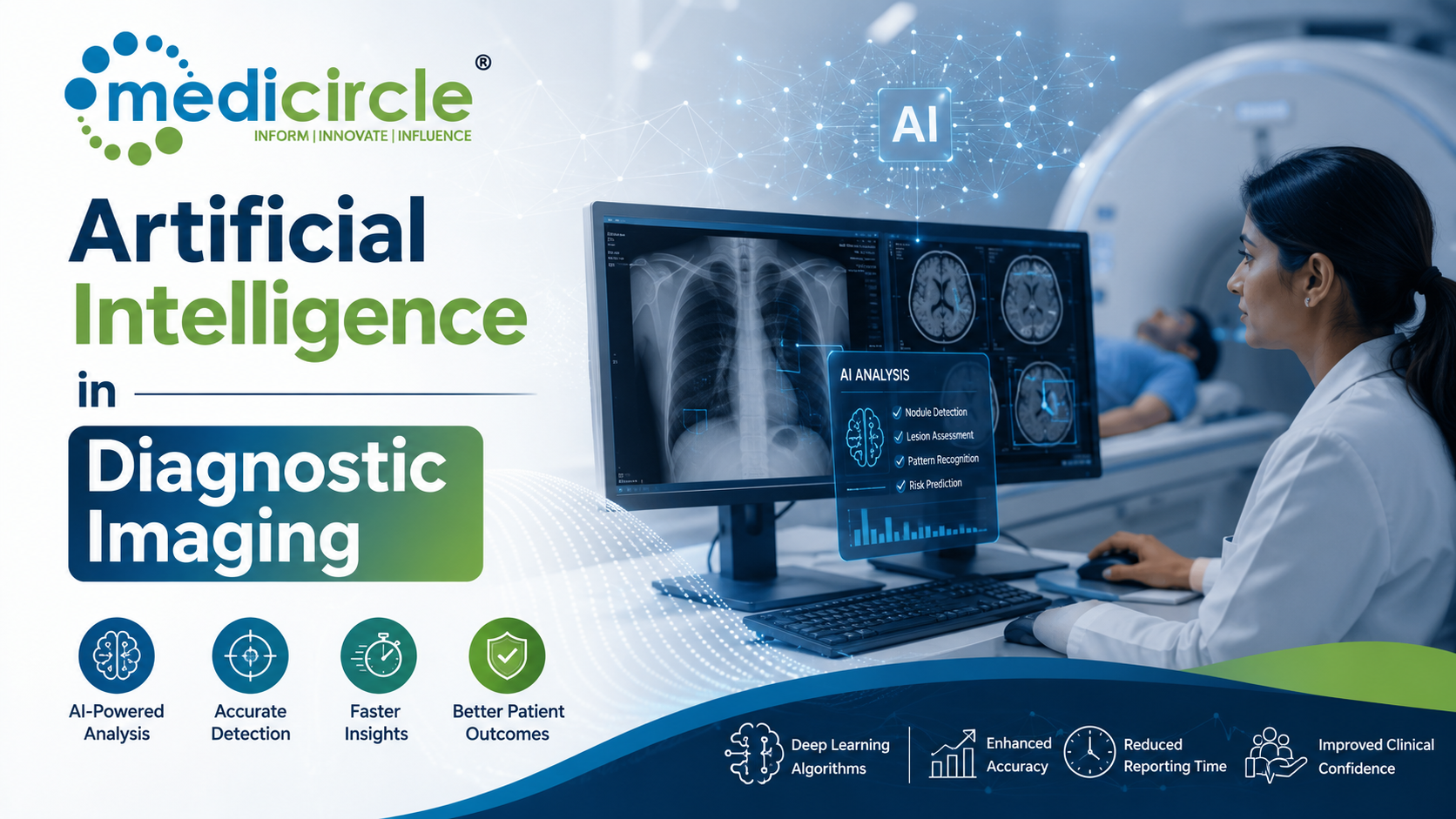
 Modern healthcare relies heavily on diagnostic imaging as its primary investigative tool. Across global health networks, the sheer volume of advanced imaging modalities—including Computed Tomography (CT), Magnetic Resonance Imaging (MRI), Digital Mammography, and Digital Radiography—has exploded.




Modern healthcare relies heavily on diagnostic imaging as its primary investigative tool. Across global health networks, the sheer volume of advanced imaging modalities—including Computed Tomography (CT), Magnetic Resonance Imaging (MRI), Digital Mammography, and Digital Radiography—has exploded.
However, this rapid growth in diagnostic data has exposed a major structural bottleneck in clinical practice: radiologist fatigue, cognitive overload, and diagnostic turnaround delays.
Interpreting a single modern multi-slice CT scan or volumetric MRI requires reviewing hundreds—sometimes thousands—of cross-sectional images under tight clinical time constraints. Prolonged exposure to high image volumes inevitably leads to visual fatigue and cognitive strain, which increases reporting variability and the risk of diagnostic errors.
Integrating Artificial Intelligence (AI) into diagnostic imaging directly resolves these operational limits. Rather than replacing human expertise, deep learning algorithms act as an advanced, continuous "second reader". AI pre-screens incoming scans, highlights subtle pathological anomalies, and streamlines clinical triage, significantly enhancing radiologist precision, minimizing diagnostic errors, and improving overall patient outcomes.
1. The Core Technological Pillars of AI-Driven Imaging
To understand how machine learning systems enhance radiologist precision, we must look at the specific algorithmic frameworks driving automated image processing:
Convolutional Neural Networks (CNNs) and Deep LearningThe core engine behind modern medical computer vision is the Convolutional Neural Network (CNN). Designed specifically to analyze visual spatial hierarchies, CNNs extract complex features directly from raw image pixels without relying on manual rules.
They analyze subtle changes in texture, density, and anatomical borders across different image slices. This enables deep learning models to identify micro-calcifications in mammograms, tiny lung nodules on CT scans, or early ischemic strokes on non-contrast head CTs far earlier than the human eye typically catches them.
Quantitative Radiomics and Non-Invasive BiomarkersAI shifts radiology from purely qualitative visual inspection to automated, high-throughput quantitative analysis. Radiomics algorithms extract thousands of mathematical data points—including voxel intensity distributions, spatial heterogeneity, and micro-surface textures—from a target lesion.
This hidden data allows AI models to differentiate benign from malignant tissues non-invasively, predict tumor genetic profiles, and assess treatment responses earlier than traditional visual reviews allow.
Generative AI and Medical Foundation ModelsBeyond single-task detection models, modern radiology is adopting multi-modal foundation models. These large-scale deep learning frameworks learn broad anatomical representations across different imaging types—such as X-rays, CTs, MRIs, and ultrasounds.
When combined with Natural Language Processing (NLP), these models help automatically generate preliminary structured reports, check drafted impression notes against visual findings, and flag inconsistencies before the final report is signed.
2. Targeted Clinical Applications: Where AI Mitigates Human Error
AI algorithms operate across specific diagnostic domains, serving as targeted safety nets against common human errors:
- Emergency Neuroimaging and Triage Priority: In acute neuro-emergencies like intracranial hemorrhage or large vessel occlusion (LVO) strokes, every minute matters. AI triage engines automatically analyze head CT scans the moment they are completed at the scanner, instantly alerting on-call neuroradiologists and stroke teams to critical findings. This automated triage drops door-to-needle times significantly, saving brain tissue.
- Oncology Screening and Micro-Lesion Detection: In high-volume screening programs—such as low-dose CT lung cancer screening or digital breast tomosynthesis—radiologists process thousands of normal scans to find a few subtle malignancies. AI models pre-screen these studies, drawing bounding boxes around micro-nodules or tissue distortions. This secondary review reduces missed lesion rates by up to 25–30%.
- Musculoskeletal Fracture Identification: Subtle hairline fractures, non-displaced scaphoid breaks, or pediatric growth-plate injuries are frequently missed on emergency X-rays due to overlapping bone structures. AI detection overlays highlight subtle cortical disruptions, giving emergency room physicians and general radiologists immediate diagnostic support.
- Automated Volumetric and Organ Segmentation: Measuring organ volumes, tracking liver fat content, or measuring complex abdominal aortic aneurysms manually is time-consuming and subject to inter-observer variability. AI algorithms segment 3D organs and lesions automatically in seconds, providing highly reproducible, objective quantitative tracking across serial follow-up scans.
3. High-Performance Action Plan for Radiology Department Directors
To successfully integrate deep learning diagnostics into clinical workflows while avoiding automation bias and integration bottlenecks, execute this multi-phase deployment roadmap:
- Execute PACS/RIS Infrastructure Integration and Data Pipeline Security
Phase 1
Secure your technology core early. Ensure your Picture Archiving and Communication System (PACS) and Radiology Information System (RIS) support DICOM-SR (Structured Reporting) and AI result overlays natively without disrupting core reading speeds. - Deploy Targeted High-Impact Algorithms and Conduct Local Validation
Phase 2
Introduce AI tools strategically. Deploy FDA-cleared algorithms for high-volume or high-risk tasks—such as stroke triage, lung nodule detection, or mammography screening—and validate their performance against your local patient demographics. - Enforce Human-in-the-Loop Safeguards and Continuous Performance Audits
Phase 3
Lock in clinical oversight. Train radiologists to treat AI outputs as supportive second opinions rather than absolute truths, avoiding automation bias, and run monthly audits to track algorithm drift and false-positive rates.
Actionable Strategy: Digital Governance and Regulatory Integration
- Link Diagnostic Reports Natively with National Digital Health Registries: Ensure all AI-assisted imaging reports, quantitative radiomic summaries, and critical finding alerts sync cleanly with unified electronic health platforms—such as the Ayushman Bharat Health Account (ABHA) network—preserving a secure, portable medical history for cross-specialty care teams.
- Coordinate Radiology Upskilling Hours Natively via Unified Academic Registries: Track all advanced artificial intelligence in medicine courses, computer vision workshops, and digital health informatics training completed by clinical staff natively using verified registries like the APAAR ID system within the Academic Bank of Credits (ABC) network to simplify credentialing audits.
- Establish Semi-Annual Algorithmic Drift and Safety Audits: Maintain continuous oversight of deployed models. Review AI performance metrics twice yearly alongside medical physicists and IT leads, tracking algorithm sensitivity, false-positive counts, and reporting turnaround speeds to keep your diagnostic infrastructure operating safely.
Frequently Asked Questions (FAQs)
Q1. Will artificial intelligence eventually replace human radiologists?No. AI functions as an advanced diagnostic assistant rather than a replacement. While AI excels at rapid pattern recognition and quantitative analysis, human radiologists are essential for contextualizing complex clinical histories, performing interventional procedures, communicating with care teams, and taking ultimate legal responsibility for diagnoses.
Q2. What is "automation bias," and why is it a risk in AI radiology workflows?Automation bias occurs when a clinician over-relies on automated AI output, uncritically accepting a false positive or overlooking an abnormality because the software failed to flag it. Robust "human-in-the-loop" training ensures radiologists use AI as a secondary reader rather than a final decision-maker.
Q3. How does AI triage improve patient outcomes in emergency stroke care?AI algorithms analyze non-contrast head CTs in seconds, instantly identifying large vessel occlusions or intracranial hemorrhages. By automatically prioritizing these critical scans at the top of the reading list and alerting stroke teams immediately, AI reduces door-to-treatment times, preserving functional brain tissue.
Q4. How does linking AI imaging reports to an ABHA ID support patient care?Integrating diagnostic reports with an ABHA ID ensures that AI-assisted findings, key image snapshots, and quantitative tracking notes are securely stored in the patient's unified digital health profile, allowing treating oncologists, surgeons, and specialists to access the data anywhere instantly.
Q5. What is the role of an APAAR ID in verifying radiologist credentials in digital health?An APAAR ID acts as a secure digital ledger that records a medical professional's verified academic accomplishments, sub-specialty fellowships, and technical AI literacy certifications across national databases, simplifying credentialing checks.
Q6. How does AI help reduce radiologist burnout in high-volume imaging centers?AI automates repetitive tasks—such as manual 3D organ measurement, image reconstruction, and preliminary draft generation. By streamlining routine work and reducing cognitive strain, AI helps lower burnout rates among clinical staff.
Q7. What is the difference between computer-aided detection (CADe) and deep learning AI models?Traditional CADe systems relied on rigid, hand-crafted rules that often generated high rates of false positives, causing alarm fatigue. Deep learning models, such as Convolutional Neural Networks, learn complex visual representations directly from massive datasets, yielding significantly higher sensitivity and specificity.
Q8. What parameters are continuously tracked on an AI radiology performance scorecard?A comprehensive department scorecard tracks metrics like algorithm sensitivity and specificity rates, false-positive frequencies per scan, PACS processing latency times, emergency triage alert-to-read intervals, and overall report turnaround times.
Q9. How fast can an imaging department expect an improvement in report turnaround times after deploying AI?When a radiology department integrates AI-driven triage and automated draft generation into its PACS environment, performance returns are immediate. Critical alert turnaround times and overall report backlogs often drop within 2 to 4 weeks of active deployment.
Q10. What immediate action should a department lead take if an AI algorithm displays a sudden drop in accuracy?The department lead must act swiftly: temporarily suspend the algorithm's active triage queue, switch the system back to standard manual reading protocols, contact the vendor and IT integration teams to investigate potential data input changes (algorithmic drift), and re-validate performance against fresh local scans before re-enabling automated features.





-23-300x250.png)

.jpeg)

.jpg)
.jpg)
.jpg)
.jpg)
.jpg)
.jpg)
.jpg)
.jpg)
.jpg)