 Monday, October 05, 2026
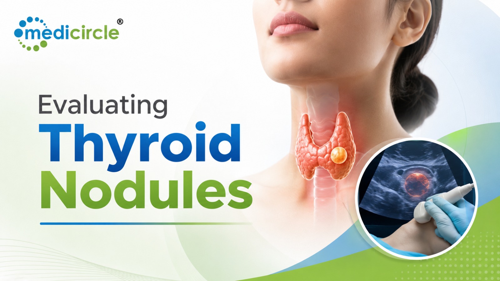
 Thyroid nodule evaluation combines ultrasound risk stratification, ultrasound-guided FNAC, and Bethesda classification to distinguish benign from malignant lesions, reduce unnecessary surgery, and guide timely treatment decisions.




Thyroid nodules are an exceptionally common clinical finding, detected in up to 50\%\text{ to }65\% of the general adult population through high-resolution ultrasound. While the vast majority (>90\%) of these lesions are benign hyperplastic or colloid nodules, the primary clinical objective is to accurately and efficiently identify the small subset that harbors clinically significant thyroid malignancy.
Over the past two decades, the diagnostic pathway for thyroid lesions has evolved from diagnostic open hemithyroidectomies to minimally invasive Fine Needle Aspiration Cytology (FNAC) standardized under The Bethesda System for Reporting Thyroid Cytopathology (TBSRTC).
When evaluating thyroid nodules, combining ultrasound risk stratification (e.g., ACR TI-RADS or EU-TIRADS) with ultrasound-guided FNAC and Bethesda classification enables clinicians to avoid unnecessary surgeries for benign conditions while ensuring timely oncological resection for high-risk carcinomas.
1. Initial Triage: Ultrasound Features and TI-RADS Stratification
Before proceeding directly to aspiration, every thyroid nodule requires comprehensive ultrasonographic evaluation. Fine Needle Aspiration is not indicated based on nodule size alone; it is dictated by the combination of maximum diameter and specific sonographic features of malignancy.
High-Risk Sonographic Features- Microcalcifications (Punctate Echogenic Foci): Strongly associated with papillary thyroid carcinoma (PTC) psammoma bodies.
- Non-Parallel Orientation ("Taller-than-Wide"): Suggests aggressive anteroposterior tissue invasion.
- Irregular, Lobulated, or Infiltrative Margins: Indicates micro-invasion into surrounding thyroid parenchyma.
- Marked Hypoechogenicity: Lesion appears darker than the adjacent strap musculature.
- Solid Composition: Solid nodules carry a significantly higher malignancy risk than purely cystic or spongiform nodules.
2. Ultrasound-Guided FNAC: Technique and Adequacy
Palpation-guided aspiration is historically associated with higher non-diagnostic rates. Modern clinical guidelines mandate real-time ultrasound-guided FNAC using fine-gauge needles (23\text{G to }27\text{G}) via capillary action (non-aspiration technique) or gentle negative pressure suction.
Specimen Adequacy CriteriaTo be classified as cytologically satisfactory (diagnostic), the specimen must contain a minimum of 6 distinct clusters of well-preserved, well-stained follicular cells, with each group comprising at least 10 cells, preferably on a single slide.
- Exception: Solid nodules with marked nuclear atypia or nodular colloid-rich lesions containing abundant thick colloid qualify as diagnostic even if the strict cell-count threshold is not met.
3. The Bethesda System for Reporting Thyroid Cytopathology
The Bethesda System provides a standardized, six-tier diagnostic reporting framework that links cytological interpretations directly to estimated malignancy risks and consensus clinical management algorithms.
- Bethesda I: Non-Diagnostic / Unsatisfactory (Risk of Malignancy: 5%–10%): Represents inadequate cellularity, obscuring blood, thick drying artifacts, or cyst fluid only. Management requires repeat ultrasound-guided FNAC after a 4-to-6-week healing window.
- Bethesda II: Benign (Risk of Malignancy: 2%–4%): Cytology shows abundant thin colloid, flat sheets of uniform follicular cells, and benign macrofollicles (e.g., adenomatoid nodule, Hashimoto's thyroiditis). Management is conservative clinical and ultrasound follow-up at 12–24 months without surgery.
- Bethesda III: Atypia of Undetermined Significance / Follicular Lesion of Undetermined Significance (AUS/FLUS) (Risk of Malignancy: ~10%–30%): Features cells with architectural or nuclear atypia that are insufficient to classify as suspicious or neoplastic. Management involves molecular testing (e.g., BRAF, RAS, RET/PTC) or repeat FNAC.
- Bethesda IV: Follicular Neoplasm / Suspicious for a Follicular Neoplasm (FN/SFN) (Risk of Malignancy: ~25%–40%): Demonstrates high cellularity composed of microfollicles or crowded cells lacking nuclear features of papillary carcinoma. Cytology cannot distinguish benign follicular adenoma from follicular carcinoma; definitive diagnosis requires surgical diagnostic lobectomy to assess capsular or vascular invasion.
- Bethesda V: Suspicious for Malignancy (SUSP) (Risk of Malignancy: ~60%–80%): Exhibits several cytological features of malignancy (such as nuclear grooves, pseudoinclusions, or prominent nucleoli) that fall just short of definitive confirmation for papillary, medullary, or anaplastic carcinoma. Management warrants surgical resection (diagnostic lobectomy or total thyroidectomy).
- Bethesda VI: Malignant (Risk of Malignancy: ~97%–100%): Unequivocal cytological diagnosis of Papillary Thyroid Carcinoma (PTC), Medullary Thyroid Carcinoma (MTC), Anaplastic Carcinoma, or Thyroid Lymphoma. Management requires definitive surgical resection (total thyroidectomy or lobectomy) with consideration of central neck dissection.
4. High-Performance Action Plan: 4-Phase Nodule Evaluation Roadmap
To execute a structured diagnostic and therapeutic workup, clinical teams can follow a systematic four-phase clinical pathway:
- Check Serum TSH and Perform Comprehensive Neck Ultrasound
Phase 1: Biochemical & Sonographic Baseline
Measure serum Thyroid Stimulating Hormone (TSH) and evaluate nodule vascularity and lymph node stations (levels II–VI). If TSH is suppressed, perform a radionuclide thyroid scintigraphy scan to assess for a hyperfunctioning ("hot") nodule. - Perform Precision Real-Time Ultrasound-Guided FNAC
Phase 2: Targeted US-Guided Aspiration
Target the solid, hypoechoic, and calcified regions of TI-RADS stratified lesions. Prepare both air-dried (Giemsa) and alcohol-fixed (Papanicolaou) smears for expert cytopathological analysis. - Assign Bethesda Classification & Consider Molecular Markers
Phase 3: Bethesda Cytology Tiering
Correlate cytological findings with Bethesda Categories I–VI. For indeterminate nodules (Bethesda III/IV), utilize molecular diagnostics (gene expression profiling or mutational panels) to rule out high-risk genetic alterations. - Execute Longitudinal Surveillance or Targeted Surgical Resection
Phase 4: Multidisciplinary Treatment Execution
Discharge Bethesda II cases to outpatient ultrasound monitoring; route Bethesda IV, V, and VI cases to head & neck endocrine surgery boards for staged lobectomy or total thyroidectomy with nerve monitoring.
Actionable Strategy: Digital Governance & Health Record Portability
- Preserve Longitudinal Endocrine Records via Digital Health Networks: Link ultrasound imaging reports, serial nodule volume measurements, and cytopathology slide reports to universal digital health accounts—such as the ABHA ID (Ayushman Bharat Health Account) pipeline. This preserves longitudinal data for multi-year surveillance and prevents duplicate biopsy procedures.
- Verify Specialist Pathology Credentials via Academic Repositories: Ensure reviewing cytopathologists, head and neck surgeons, and endocrinologists hold certified qualifications verified through national digital registries like the APAAR ID system within the Academic Bank of Credits (ABC) network.
- Track Calcitonin Biomarkers for Medullary Screening: In family-inherited or atypical nodule presentations, cross-reference serum calcitonin and carcinoembryonic antigen (CEA) levels with cytological findings to detect early Medullary Thyroid Carcinoma (MTC).
Frequently Asked Questions (FAQs)
Q1. What is the main purpose of performing Fine Needle Aspiration Cytology (FNAC) on a thyroid nodule?The main purpose of FNAC is to obtain a representative cellular sample from a suspicious thyroid nodule to determine whether it is benign, indeterminate, or malignant, allowing clinicians to make evidence-based surgical or surveillance decisions.
Q2. Why is ultrasound guidance essential during thyroid FNAC?Real-time ultrasound guidance ensures the needle tip is positioned precisely within the suspicious solid or calcified areas of the nodule while avoiding purely cystic or necrotic fluid pockets, significantly reducing non-diagnostic rates.
Q3. What is the difference between a "hot" and "cold" thyroid nodule?A "hot" (hyperfunctioning) nodule absorbs more radioactive tracer than normal thyroid tissue on a scintigraphy scan and is almost always benign (<1\% malignancy risk). A "cold" (non-functioning) nodule absorbs little to no tracer and carries a higher risk of malignancy, warranting FNAC if sonographically suspicious.
Q4. Can FNAC differentiate between a benign follicular adenoma and follicular carcinoma?No. Cytology only evaluates individual cells or small groups; it cannot assess architectural features like capsular invasion or vascular invasion. Differentiating follicular adenoma from follicular carcinoma requires histopathological evaluation of the entire excised surgical specimen (Bethesda IV).
Q5. What is the recommended management for a Bethesda III (AUS/FLUS) nodule?Current guidelines recommend either repeating the ultrasound-guided FNAC after a few weeks, utilizing advanced molecular testing (to check for gene mutations like BRAF or RAS), or proceeding with a diagnostic lobectomy depending on clinical risk factors.
Q6. How often should a confirmed benign (Bethesda II) nodule be monitored?Benign nodules are typically monitored with clinical examination and repeat thyroid ultrasound every 12 to 24 months. If the nodule remains stable in size and appearance, the surveillance interval can be extended.
Q7. What are psammoma bodies, and why are they significant in thyroid cytology?Psammoma bodies are microscopic, concentric laminated calcifications. When identified in thyroid cytology, they are a strong diagnostic indicator of Papillary Thyroid Carcinoma (PTC).
Q8. Is a larger thyroid nodule always more likely to be cancerous?Not necessarily. While size determines whether a nodule meets the threshold for biopsy, the intrinsic sonographic architecture (composition, echogenicity, shape, margins, and calcifications) is a more accurate predictor of malignancy than size alone.
Q9. What complications are associated with thyroid FNAC?Thyroid FNAC is a safe, minimally invasive outpatient procedure. Minor complications include localized discomfort, mild bruising, and temporary swelling. Significant hematomas, infections, or recurrent laryngeal nerve injuries are rare.
Q10. What is the role of molecular testing in thyroid nodule evaluation?Molecular testing analyzes aspirated cells for specific gene mutations (e.g., BRAF V600E, TERT, RET/PTC) and gene expression profiles. It provides diagnostic clarity for indeterminate Bethesda III and IV categories, helping patients avoid diagnostic surgery if the molecular profile is benign.






.jpeg)
.jpg)
.jpg)
.jpg)
.jpg)
.jpg)
.jpg)








.jpeg)
.jpeg)


.png)