 Monday, October 05, 2026
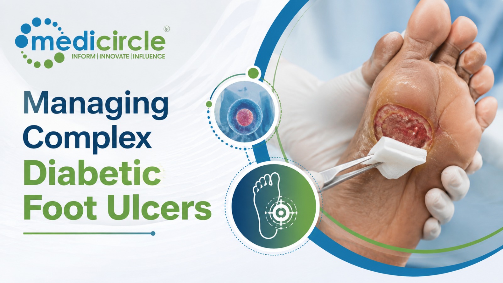
 Diabetic Foot Ulcers (DFUs) represent one of the most severe, costly, and debilitating complications of Diabetes Mellitus. Driven by a triad of peripheral neuropathy, micro/macrovascular ischemia, and impaired immune response, a simple blister or minor trauma can rapidly progress to deep tissue necrosis, osteomyelitis, and lower-extremity amputation




Diabetic Foot Ulcers (DFUs) represent one of the most severe, costly, and debilitating complications of Diabetes Mellitus. Driven by a triad of peripheral neuropathy, micro/macrovascular ischemia, and impaired immune response, a simple blister or minor trauma can rapidly progress to deep tissue necrosis, osteomyelitis, and lower-extremity amputation.
Managing complex DFUs requires moving away from isolated wound care toward an integrated, limb-salvage framework centered on three core pillars: Multidisciplinary Wound Care & Debridement, Biomechanical Offloading, and Vascular Assessment & Revascularization.
1. Clinical Etiology & Diagnostic Triage
Complex DFUs rarely stem from a single cause. Accurate clinical classification guides immediate therapeutic priorities:
Neuropathic Ulcers- Mechanism: Loss of protective sensation (LOPS) due to distal symmetric sensorimotor polyneuropathy. Unnoticed repetitive mechanical stress creates hyperkeratotic calluses, tissue breakdown, and painless ulceration—typically over plantar weight-bearing areas (metatarsal heads).
- Clinical Presentation: Warm foot, bounding pulses, dry/cracked skin (autonomic neuropathy), painless ulcer surrounded by heavy callus.
- Mechanism: Combined neuropathic sensory loss and peripheral artery disease (PAD), frequently aggravated by microvascular dysfunction and medial arterial calcification (Monckeberg's sclerosis).
- Clinical Presentation: Cold, pale, or cyanotic foot; diminished or absent dorsalis pedis/posterior tibial pulses; delayed capillary refill; painful or nocturnal resting pain (if sensory nerves remain partially intact); ulcers positioned on distal toes, lateral foot margins, or heel margins.
2. The Three Pillars of Advanced DFU Management
Successful DFU management and limb salvage rely on a structured, three-pronged approach:
Pillar 1: Surgical Debridement and Advanced Wound Bed PreparationThe primary goal of debridement is converting a chronic, stagnant wound into an active, healing acute wound:
- Sharp Surgical Debridement: The gold standard. Removes necrotic tissue, senescent fibroblasts, dense bacterial biofilms, and hyperkeratotic callus margins until viable, bleeding tissue is exposed.
- Infection Control & Biofilm Management: Clinical diagnosis of infection relies on local inflammatory signs (rubor, calor, dolor, purulence) rather than surface swabs. Systemic empirical antibiotics are reserved for active clinical infections (classified via IWGDF/IDSA guidelines), targeting common pathogens including Staphylococcus aureus, Streptococci, and anaerobes in deep tissue involvement.
- Advanced Therapies: Utilizing Negative Pressure Wound Therapy (NPWT) to stimulate granulation tissue, hyperbaric oxygen therapy (HBOT) for refractory hypoxic wounds, and bio-engineered cellular/acellular dermal matrices.
Without eliminating mechanical pressure and shear stress over the ulcer site, advanced topical therapies and surgical procedures fail.
- Total Contact Casting (TCC): The gold standard for offloading plantar neuropathic ulcers. TCC distributes body weight across the entire plantar surface and lower leg, preventing localized shear forces. It enforces patient compliance while reducing peak plantar pressure by up to 80–90%.
- Removable Cast Walkers (RCWs): Effective alternative when TCC is contraindicated (e.g., active deep infection, heavy exudate, or severe arterial ischemia), provided patient compliance is strictly maintained.
- Custom Orthotics & Post-Healing Footwear: Specialized depth shoes with rigid rocker soles and custom molded insoles are mandatory post-closure to prevent ulcer recurrence.
Ischemia is the single greatest predictor of delayed healing and major amputation. Every complex DFU must undergo early vascular evaluation:
- Non-Invasive Vascular Testing: Ankle-Brachial Index (ABI) can be falsely elevated due to non-compressible calcified vessels in diabetic patients. Therefore, Toe Brachial Index (TBI), Toe Systolic Pressure, and Transcutaneous Oximetry (\text{TcPO}_2) serve as superior metrics for tissue perfusion.
- Angiography & Revascularization: Indicated when \text{TcPO}_2 < 30\text{ mmHg}, toe pressure < 30\text{ mmHg}, or when an ulcer fails to reduce in size by >50\% after 4 weeks of optimal wound care.
- Revascularization Modalities: Combines endovascular techniques (angioplasty, stenting, atherectomy) and open bypass surgery (using autologous vein grafts) to restore direct inline blood flow through at least one major infrapopliteal artery to the specific anatomical angiosome of the ulcer.
3. Structural Comparison: Standard Care vs. Multidisciplinary Limb-Salvage Team
Comparing fragmented care with an integrated multidisciplinary team highlights how structured care improves clinical outcomes:
- Diagnostic Timeline: Fragmented care delays vascular testing and deep infection detection. Multidisciplinary teams perform immediate bedside sensory, vascular (\text{TBI/TcPO}_2), and wound-bed audits on day one.
- Offloading Approach: Standard care relies on soft surgical shoes or custom flats, achieving suboptimal pressure reduction. Multidisciplinary teams deploy Total Contact Casts (TCC) or rigid Removable Cast Walkers (RCWs), lowering plantar pressure by up to 90%.
- Revascularization Timing: Ad-hoc care refers patients to vascular specialists only after months of failed conservative wound care. Integrated teams trigger immediate vascular imaging and angioplasty/bypass within days if ischemic parameters are breached.
- Infection & Bone Management: Fragmented care relies on prolonged oral antibiotics without surgical debridement. Multidisciplinary care executes sharp debridement, targeted tissue cultures, MRI evaluation for osteomyelitis, and antimicrobial stewardship.
- Clinical Outcomes: Ad-hoc care leads to high ulcer recurrence rates and elevated risk of major below-knee/above-knee amputations. Multidisciplinary care accelerates healing times, minimizes major amputation rates, and lowers long-term healthcare costs.
4. High-Performance Action Plan for Clinical Wound Teams
To establish an effective limb-salvage workflow for complex diabetic foot ulcers, clinical teams can follow this three-phase operational plan:
- Execute Immediate Vascular, Neuropathic, and Infection Audits
Phase 1: Triage & Vascular Assessment
Screen for sensory loss using a 10g Semmes-Weinstein monofilament. Measure TBI or \text{TcPO}_2 to rule out severe peripheral artery disease. Probe the ulcer to bone (PTB test) and order plain radiographs or MRI if deep infection or osteomyelitis is suspected. - Perform Sharp Debridement and Apply Total Contact Casting
Phase 2: Surgical Debridement & Offloading
Remove all devitalized tissue, hyperkeratotic callus, and biofilm until bleeding wound margins are achieved. Apply a Total Contact Cast (TCC) or rigid non-removable cast walker for uninfected neuropathic plantar ulcers. - Trigger Early Angiography and Post-Closure Footwear Protocols
Phase 3: Revascularization & Maintenance
If tissue perfusion is compromised (\text{TcPO}_2 < 30\text{ mmHg} or TBI < 0.7), schedule urgent endovascular or open revascularization. Upon full epithelialization, transition the patient into custom-molded orthotics and depth footwear to prevent recurrence.
Frequently Asked Questions (FAQs)
Q1. What is the semmes-weinstein monofilament test, and how is it used in DFU screening?The 10g Semmes-Weinstein monofilament test is a non-invasive diagnostic tool used to assess loss of protective sensation (LOPS). Applying the filament to specific plantar sites until it buckles identifies patients with sensory neuropathy who are at high risk for painless foot ulceration.
Q2. Why is the Ankle-Brachial Index (ABI) often unreliable in diabetic patients?Diabetic patients frequently develop medial arterial calcification (Mönckeberg's arteriosclerosis), making peripheral lower-leg arteries stiff and non-compressible. This yields falsely elevated or normal ABI values (>1.3) despite severe underlying distal ischemia. The Toe-Brachial Index (TBI) is preferred because digital arteries are rarely affected by severe medial calcification.
Q3. What is the "Probe-to-Bone" (PTB) test?The PTB test involves gently probing the base of an open ulcer with a sterile metallic blunt-tipped probe. If the probe contacts a hard, gritty bone surface, there is a high positive predictive value for underlying osteomyelitis, warranting further imaging (MRI) and deep tissue cultures.
Q4. When is Total Contact Casting (TCC) contraindicated?TCC is contraindicated in the presence of unmanaged deep tissue infection, active osteomyelitis, severe peripheral artery disease (where rigid compression can worsen tissue ischemia), unhealed surgical incisions, or heavily exudative wounds requiring daily dressing changes.
Q5. What is the role of hyperbaric oxygen therapy (HBOT) in DFU management?HBOT involves breathing 100% oxygen at elevated atmospheric pressure. It increases dissolved plasma oxygen concentration, enhancing fibroblast proliferation, collagen synthesis, angiogenesis, and leukocyte bacterial killing in chronic, hypoxic, non-healing neuroischemic ulcers.
Q6. How does angiosome-targeted revascularization improve wound healing?The angiosome concept divides the foot into distinct anatomical regions supplied by specific source arteries (e.g., anterior tibial, posterior tibial, or peroneal arteries). Revascularizing the specific artery feeding the tissue block containing the ulcer maximizes local perfusion and accelerates healing compared to non-targeted revascularization.
Q7. What are the signs of a Charcot Neuroarthropathy flare-up in a diabetic foot?Acute Charcot foot manifests as a warm, erythematous, edematous foot with a temperature difference of >2^\circ\text{C} compared to the contralateral foot, often following minor trauma in a neuropathic patient. Radiographs may reveal bone fragmentation, joint dislocation, and arch collapse (rocker-bottom deformity).
Q8. How frequently should sharp surgical debridement be performed on a chronic DFU?Sharp surgical debridement is typically performed weekly or bi-weekly during active wound care to continuously disrupt bacterial biofilm formation, remove hyperkeratotic callus buildup, and maintain an active, granulating wound bed.
Q9. What proportion of diabetic foot ulcers recur after complete healing?Recurrence rates for DFUs are high, with roughly 40% of patients experiencing ulcer recurrence within 1 year and up to 65% within 3 years post-closure. This highlights the vital need for lifelong custom therapeutic footwear and continuous podiatric surveillance.
Q10. What immediate step should a clinician take when presenting with an infected, ischemic DFU?Admit the patient for urgent IV antibiotic therapy, order immediate non-invasive vascular imaging (TBI/duplex ultrasound) to plan rapid revascularization, perform urgent surgical drainage/debridement of infected tissue, and initiate strict non-weight-bearing offloading.






.jpeg)
.jpg)
.jpg)
.jpg)
.jpg)
.jpg)
.jpg)








.jpeg)

.jpeg)


.png)