Tuesday, October 06, 2026
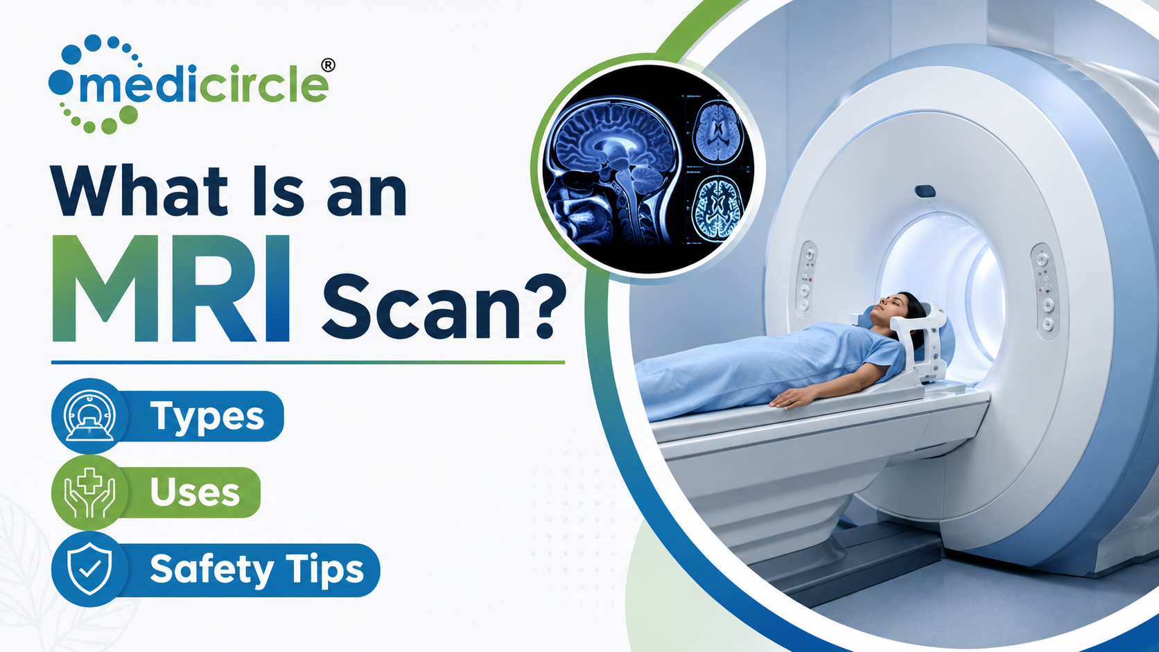
 This article explains what an MRI scan is, how it works, its clinical applications across specialties, safety considerations, preparation steps, and its expanding role in India's evolving healthcare landscape.




Introduction
Medical imaging has transformed the way doctors understand what happens inside the human body. Among the most powerful and widely used diagnostic tools today, the MRI scan stands out for its ability to produce highly detailed images of soft tissues, organs, the brain, and the spinal cord without exposing the patient to any radiation. In India, demand for MRI scans has grown substantially over the past decade, with an increasing number of hospitals in both metro cities and Tier 2 towns offering this technology as part of comprehensive diagnostic services.
For many patients, the experience of being asked to undergo an MRI scan raises a host of questions. What exactly does an MRI scan detect? Is it safe? How should one prepare? What is the difference between this scan and a CT scan? These are important questions, and understanding the answers helps patients make more informed decisions about their healthcare.
This article provides a thorough, evidence-based guide to what an MRI scan is, how it works, what it can diagnose, what patients should expect during the process, and what safety considerations apply.
Understanding Magnetic Resonance Imaging: The Basics
MRI stands for Magnetic Resonance Imaging. It is a non-invasive diagnostic test that uses a combination of a powerful magnetic field, radio waves, and computer processing to generate detailed cross-sectional images of the internal structures of the body.
Unlike X-rays and CT scans, an MRI scan does not use ionizing radiation. Instead, it works by detecting the behavior of hydrogen atoms present in water and fat throughout the body. When a patient lies inside the MRI machine, the strong magnetic field causes these hydrogen atoms to align in a particular direction. A pulse of radio waves is then directed at the body, which momentarily disrupts this alignment. As the atoms return to their original position, they emit radio signals that are picked up by the machine and converted by a computer into high-resolution images.
The resulting images can show organs, muscles, blood vessels, ligaments, and even microscopic tissue changes with a level of clarity that surpasses what conventional X-rays or CT scans can provide, particularly for soft tissues and neurological structures. This makes MRI an indispensable tool for diagnosing a wide range of conditions affecting the brain, spine, joints, heart, abdomen, and pelvis.
Types of MRI MachinesThere are two primary types of MRI machines available in India and across the world:
- Closed-bore MRI: This is the traditional cylindrical machine that produces the highest quality images. The patient lies on a table that slides into the centre of the tube. The enclosed design can cause discomfort or anxiety in people who have claustrophobia.
- Open MRI: Designed with open sides to reduce the feeling of enclosure, open MRI machines are better suited for patients who are anxious, very large, or have movement difficulties. However, the image quality from open machines is generally lower than that from closed-bore systems.
In addition to these standard designs, there are also specialized MRI configurations such as the 3 Tesla (3T) MRI, which uses a stronger magnetic field than the more common 1.5T machines and provides even finer image detail, particularly for neurological and musculoskeletal assessments.
What Does an MRI Scan Detect? Clinical Applications
The range of conditions that an MRI scan can detect or monitor is extensive. Physicians across specialties rely on MRI because it provides information that no other imaging test can replicate with the same degree of soft tissue contrast.
Brain and Neurological ConditionsMRI is the imaging test of choice for most neurological conditions. It can detect brain tumors, aneurysms, strokes, white matter diseases such as multiple sclerosis, epilepsy, dementia, and traumatic brain injuries. In India, where the burden of cerebrovascular disease and neurological disorders is rising sharply, brain MRI has become one of the most requested scans in tertiary hospitals. A specialized form called functional MRI (fMRI) can also map brain activity and blood flow, which is valuable for pre-surgical planning.
Spine and Spinal CordSpinal MRI is commonly used to evaluate disc herniation, nerve compression, spinal stenosis, infections such as spinal tuberculosis (which remains prevalent in India), spinal tumors, and injuries to the spinal cord. Many patients presenting with chronic low back pain or radiating leg pain in Indian clinical settings are referred for a lumbar spine MRI as a standard part of evaluation.
Joints and Musculoskeletal SystemOrthopedic specialists frequently request MRI scans of the knee, shoulder, hip, and ankle to assess ligament tears, cartilage damage, tendon injuries, stress fractures, bone infections (osteomyelitis), and bone tumors. The ability to visualize soft tissue damage in joints without surgical intervention makes MRI particularly valuable.
Heart and Blood VesselsCardiac MRI provides detailed information about the structure and function of the heart, including chamber size, wall motion, the extent of damage after a heart attack, and congenital heart defects. It is also used to assess diseases of the aorta and other major blood vessels.
Abdominal and Pelvic OrgansMRI is highly effective at evaluating the liver, kidneys, pancreas, spleen, and bile ducts. It is frequently used to characterize liver lesions, assess inflammatory bowel disease, detect pancreatic cancer, and evaluate the uterus, ovaries, and prostate gland.
Breast ImagingIn women at high risk for breast cancer, or those with dense breast tissue where mammography may not be adequate, MRI of the breast is used as an additional screening or diagnostic tool.
What Is MRI with Contrast and When Is It Used?
A standard MRI scan can be performed without any injected medication. However, for certain clinical conditions, doctors recommend an MRI with contrast. In this procedure, a substance called gadolinium is injected intravenously before or during the scan. Gadolinium alters the magnetic properties of nearby water molecules in the body, which enhances the visibility of blood vessels, inflammation, tumors, and infection on the images.
MRI contrast is commonly used when evaluating brain tumors, assessing the extent of multiple sclerosis lesions, detecting active infection, or evaluating blood vessel abnormalities. It is generally considered safe. However, patients with severe kidney disease are at risk for a rare complication called nephrogenic systemic fibrosis if gadolinium-based contrast is used, and this needs to be assessed carefully before the procedure.
Patients are advised to inform their doctor about any prior allergic reactions to contrast agents, any kidney problems, pregnancy, or breastfeeding status before a contrast-enhanced MRI is planned.
Who Should Not Undergo an MRI Scan?
MRI is safe for the vast majority of patients when appropriate screening is carried out beforehand. However, certain situations require caution or may contraindicate the procedure:
- Patients with certain types of metallic implants, such as older pacemakers, cochlear implants, aneurysm clips, certain heart valves, or metallic fragments in the eye
- Patients who have deep-brain stimulators, spinal cord stimulators, or implanted drug pumps
- Pregnant women, particularly in the first trimester, unless the scan is clinically urgent
- Patients with severe renal failure, if gadolinium contrast is being considered
It is important to inform the referring doctor and the radiology team about all implants, prior surgeries, and metal fragments in the body before the MRI is scheduled. Modern MRI facilities in India carry out thorough pre-scan screening questionnaires to identify any risk factors.
Patients with claustrophobia can discuss their anxiety with their treating physician. Mild sedation is sometimes prescribed to help the patient remain calm during the scan, and open MRI machines may be an appropriate alternative.
How to Prepare for an MRI Scan
Preparation for an MRI scan is straightforward for most patients.
- Patients can usually eat, drink, and take their regular medications as normal, unless instructed otherwise for specialized scans
- All jewelry, watches, hairpins, eyeglasses, hearing aids, and body piercings must be removed before entering the MRI room, as metal objects interact with the magnetic field
- Patients are provided with a hospital gown to change into
- The MRI room is kept free of all metal objects for safety, which means mobile phones, credit cards, and keys must be left outside
- If contrast dye is being used, an intravenous line will be inserted into a vein in the hand or arm before the scan begins
In India, many hospitals and diagnostic centers offer prior appointment slots for MRI scans. Patients are advised to arrive early to complete the necessary paperwork and screening questionnaire.
What to Expect During and After an MRI Scan
The MRI scan itself is a painless procedure. The patient lies on a motorized table that slides into the centre of the machine. During the scan, a series of loud thumping and clicking sounds is produced as the machine generates magnetic pulses. Earplugs or headphones are provided to reduce noise discomfort.
It is essential to remain as still as possible throughout the scan, as any movement can blur the images. For certain body parts, the patient may be asked to hold their breath briefly. The technologist monitors the patient from an adjacent room through a window and maintains two-way communication through an intercom system. A call button is also provided so the patient can alert the technologist if they feel uncomfortable.
After the scan is complete, patients who have not received sedation can resume their normal activities immediately. If sedation was used, the patient will need to rest briefly and arrange for someone to drive them home.
A specialist called a radiologist analyzes the MRI images and sends a written report to the referring doctor, who will discuss the findings and recommend next steps.
MRI Scanning in the Indian Healthcare Context
Access to MRI technology in India has expanded considerably in recent years. Major public hospitals under government initiatives, including those affiliated with the Ayushman Bharat Pradhan Mantri Jan Arogya Yojana (PM-JAY) scheme, are increasingly including MRI scanning among covered diagnostic services, making this technology accessible to patients from economically weaker backgrounds who were previously unable to afford it.
Large corporate hospital chains in cities like Mumbai, Delhi, Bengaluru, Hyderabad, and Chennai now operate 3T MRI machines and offer advanced MRI-guided procedures. At the same time, standalone diagnostic centers in Tier 2 cities such as Kota, Nagpur, Bhopal, and Coimbatore have brought 1.5T MRI access closer to patients in smaller towns.
The National Health Authority's digital health initiatives, including the Ayushman Bharat Digital Mission (ABDM), are also facilitating the digitization of imaging reports and enabling patients to store and share their MRI records through their Ayushman Bharat Health Account (ABHA). This is a significant step toward creating continuity of diagnostic care across India's fragmented healthcare ecosystem.
Frequently Asked Questions
Q1: Is an MRI scan painful?
An MRI scan is generally painless. The procedure requires the patient to lie still inside the machine for 30 to 60 minutes. Some people experience mild discomfort due to the enclosed space or the noise produced by the machine. If contrast dye is used, a small injection is given intravenously, which may cause brief, minor discomfort.
Q2: Can a person with a pacemaker undergo an MRI scan?
Many newer pacemaker models are MRI-compatible, but older pacemakers may be affected by the strong magnetic field. It is essential to inform the radiologist and treating doctor about any pacemaker or implanted electronic device before scheduling an MRI. An alternative imaging test, such as a CT scan, may be recommended if an MRI is not safe.
Q3: How long does an MRI scan take?
A standard MRI scan typically takes between 30 and 60 minutes, depending on the body part being examined and whether contrast material is used. Some specialized scans, such as a full-body MRI or a cardiac MRI, may take longer. The radiologist or technologist will provide an estimated time before the procedure begins.
Q4: What is the difference between an MRI scan and a CT scan?
An MRI scan uses strong magnetic fields and radio waves to create detailed images of soft tissues, organs, and nerves without using radiation. A CT scan uses X-rays and is better suited for imaging bones and detecting internal bleeding quickly. MRI provides superior contrast for soft tissue evaluation, while CT scans are faster and more widely available in smaller hospitals across India.
Q5: Is an MRI scan safe during pregnancy?
MRI is generally considered safer than CT scanning during pregnancy because it does not use ionizing radiation. However, healthcare providers usually avoid MRI scans during the first trimester unless it is clinically urgent. Gadolinium-based contrast agents are typically not administered to pregnant women unless absolutely necessary. Pregnant women should always consult their doctor before undergoing any imaging procedure.






.jpeg)
.jpg)
.jpg)
.jpg)
.jpg)
.jpg)
.jpg)


.jpg)
.jpg)
.jpg)
.jpg)
.jpg)
.jpg)